
An 80 year-old woman enjoying good past health presented to medical ward for fever and vomiting.
GCS at presentation is 15/15. One day later, she developed recurrent generalized tonic-clonic convulsions, and GCS dropped to E1V2M4. After endotracheal intubation, CT brain was performed. ICU team was consulted for clinical impression of CVA.
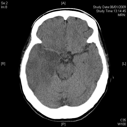
Is it CVA? Is it MCA infarct?
Click “read more” for answer
This patient suffered from herpes simplex encephalitis
On first glance, this CT brain showed feature of right MCA infarct with the right MCA being contrasted prominently, mimicking dense MCA sign. However, patient presented with fever, and recurrent seizure will be much more commonly seen in encephalitis. HSV encephalitis differerd from MCA infarct by its relatve sparing of frontal lobe, which will be inevitably involved more or less the same as temporal lobe in MCA infarct. Besides, HSV encephalitis will demontrate far less mass effect than what will be expected from the size of lesion. Hemorrhagic transformation will be rather uncommon.
Sometimes unilateral involvement is observed, making it difficult to differentiate from a tumor with vasogenic oedema. Brain tumor will be less homogenous on CT, and the oedema will be more affected at white matter. More mass effect will be expected if the lesion of this size is due to a tumor.
The diagnosis is confirmed by CSF PCR for HSV.