
Submitted by Dr HO Chun Ming Chris on 14 April, 2009
ICU, North District Hospital, Hong Kong
HKSCCM Chief Editor’s notes for overseas readers: The North District Hospital is situated near the Hong Kong-mainland China border. It is contributing a lot to serve many Hong Kong citizens who fall sick in mainland China but would like to return to Hong Kong to continue medical treatment in a Hong Kong Hospital.
This 73-year old man had perforated acute appendicitis with gangrenous caecum and peritoneal soiling. He had undergone right hemicolectomy with bowel anastomosis and appendicectomy in mainland China, but discharged himself against medical advice on post-operative day 3 and attended North District Hospital, Hong Kong, complaining of abdominal pain and respiratory distress. Clinical examination revealed fever of 38.2 degrees Celsius together with typical peritoneal signs. Bilateral abdominal drains were still in-situ with trace amount of serous drainage. The patient was admitted into our ICU and intubated for respiratory failure. Piperacillin-tazobactam (Tazocin or Zosyn; Wyeth) was started empirically.
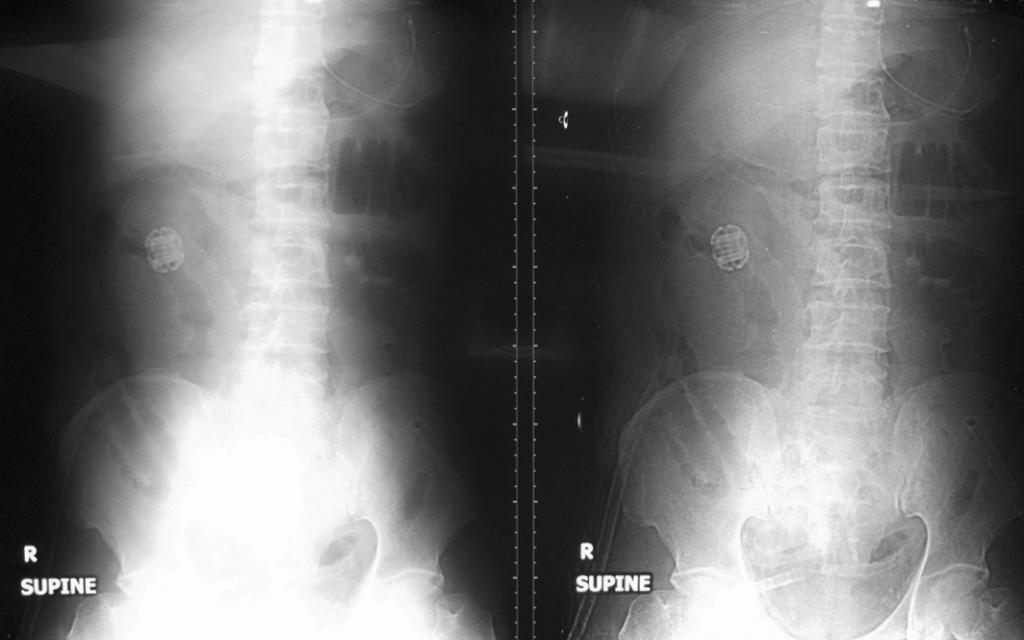
Fig. Abdominal X-ray
Please comment on the AXR’s. Click Read More to see the answer
Answer
The object seen on the R side of the abdomen is a type of Compression Anastomosis device used for intestinal anastomosis. It is not a retained surgical equipment left behind after the previous laparotomy in China as was once our suspicion.
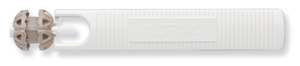
Figure. The Biofragmentable Anastomotic Ring (BAR)
This particular type is a biofragmentable anastomotic ring (BAR). As its name suggests, with time it will fragment and be expelled from the body. Problems of this device include tissue necrosis at the anastomotic site causing anastomosis dehiscence. The biofragmentation process can also be problematic and can cause bowel obstruction or strictures. It is not available in Hong Kong.
CT abdomen at the time showed diffuse mesenteric inflammatory changes consistent with postoperative changes, but no suggestion of anastomotic leakage or collections. The surgical diagnosis for this patient at the time was post-operative ileus.
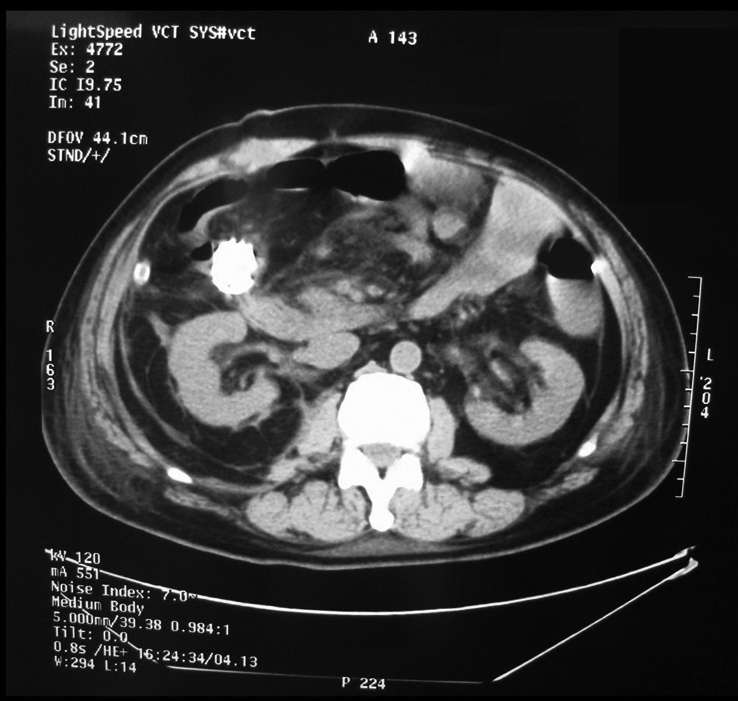
Fig. CT abdomen
However, the patient developed intra-abdominal abscess and anastomotic leakage and pus started draining from the abdominal drain. A 5-mm defect was found with pus around the anastomosis at laparotomy. The anastomosis was taken down and brought out with a double barrel enterostomy. Coagulase-negative Staphylococcus, enterococcus and candida albicans were isolated and treated accordingly. The patient survived.