
Submitted by Dr Chan Ka Hing Jacky, ICU, Tseung Kwan O Hospital, on 15 August 2009
A 64-year-old male with history of gout presented to AED for on and off chest discomfort for 10 days. He had an episode of syncope with suspected head injury at home. Blood pressure was 103/76 and pulse rate was 126/min. Oxygen saturation was 96% room air and respiratory rate was 16/min. His GCS was 15/15 and he was afebrile. Two ECGs were done in AED and they were shown below. CXR was clear.
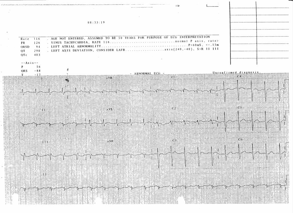
1st ECG
1st ECG (click image to enlarge)

He developed generalized tonic clonic convulsion followed by cardiac arrest in AED. CPR was started immediately and he regained spontaneous circulation about 20 minutes later. He was intubated and put on high dose vasopressors because of shock. His GCS remained 3/15 after resuscitation. He showed some neurological recovery afterwards and was then transferred to ICU for close monitoring and further management.
Bedside echocardiogram in ICU showed moderate TR with TRPG 30mmHg. RA and RV were dilated and there was mild RV hypokinesia. LVEF was satisfactory and there was no regional wall mechanical abnormality. Serum troponin T level was 0.17. CT brain (plain) and CT thorax with contrast were subsequently performed.
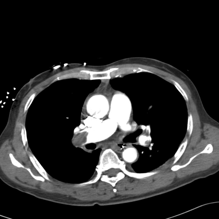
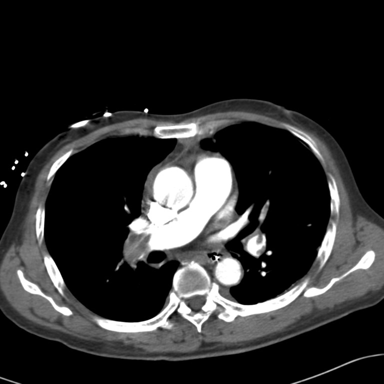
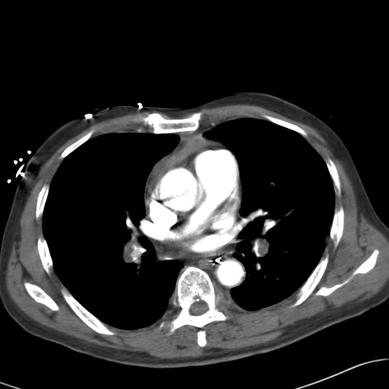
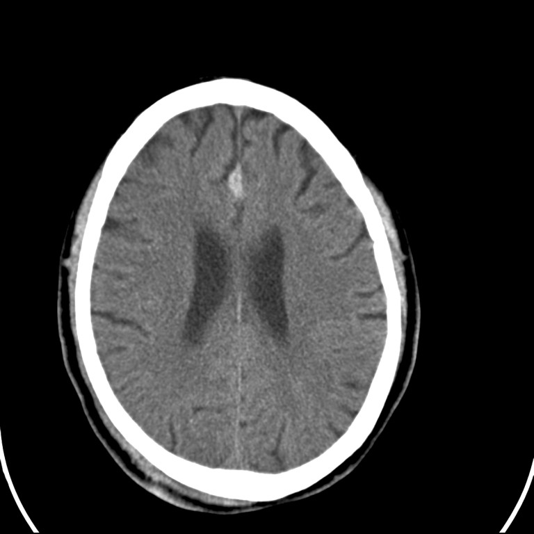
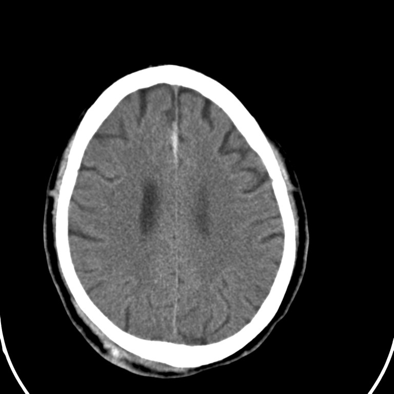
What abnormalities are shown?
1st ECG showed sinus tachycardia, Q-wave and inverted T-wave over lead III. 2nd ECG done 15 minutes later showed RBBB also. Together with the echocardiogram findings, the most likely cause for cardiac arrest and hypotension would be massive pulmonary embolism. This was confirmed by CT thorax, which revealed extensive bilateral filling defects at the main pulmonary arteries. Filling defect in left popliteal vein was also noted. CT brain however showed acute subdural haemorrhage.
Cardiothoracic unit of Queen Elizabeth Hospital was consulted. It was suggested to consult neurosurgical unit for fitness of anticoagulation as it was required even in surgical embolectomy. CT brain was repeated as suggested by neurosurgical unit and it showed no interval change. In view of the life-threatening condition and repeat CT brain result, neurosurgeon suggested proceeding to anticoagulation. Cardiothoracic surgeon was reconsulted again and surgical embolectomy was not available within short period of time. After pros and cons of anticoagulation were discussed with patient’s relatives, heparin infusion was started with close monitoring of neurological state. Tight heparin infusion regime was adopted with aPTT targeting at 45-50 sec. Unfractionated heparin was used because it had a shorter half-life than low molecular weight heparin, thus allowing rapid titration of doses. Antidote such as protamine sulphate was also available to reverse its effect in case bleeding occurred.
Vasopressor support was subsequently weaned off and patient was extubated. Repeat CT brain several days revealed no interval change for subdural haemorrhage. Patient was transferred to general ward and low molecular weight heparin was initiated after heparin infusion stopped. Malignancy screening and lifelong warfarin were decided after haematologist assessment. MRI brain was also arranged to investigate possible cause for acute subdural haemorrhage. Patient was discharged home after an uneventful stay in ward and would be followed up in outpatient department.
Discussion
Management of pulmonary embolism in critically ill can be categorized into three aspects: 1. Assessment of right ventricular function. 2. Right ventricular resuscitation. 3. Relieving pulmonary artery obstructing thrombus.
Assessment of RV function can be achieved by imaging, laboratory tests or combined methods. Imaging techniques include echocardiography and contrast enhanced multi-detector helical CT. These can identify those patients without RV dilation at lower risk of complications and mortality. Serum troponin may be elevated early but may not persist despite persistent RV dysfunction. B-type natriuretic peptide (BNP) and N-terminal pro-BNP, which are indicators of increased cardiac wall stress and myocardial hypoxia, may also be raised. Both echocardiogram and serum troponin test were performed in our case.
Right ventricular resuscitation includes volume administration, vasopressors and vasodilators. Judicious volume infusion improves cardiac output whereas overdistention of RV impairs coronary perfusion and LV filling, decreasing LV output. Nor-adrenaline is superior to phenylephrine in increasing cardiac output and RV coronary blood flow. Inhaled nitric oxide and sidenafil can also be used to lower pulmonary arterial pressure and unload RV. Fluid challenge and nor-adrenaline infusion were used to resuscitate our patient.
Ways to relieve pulmonary artery obstructing thrombus consist of thrombolytic therapy, anticoagulation, percutaneous catheter devices and surgical embolectomy. In our case, thrombolytic was contra-indicated because of acute subdural haemorrhage but surgical intervention was not available. We had to strike a balance between treating this life-threatening condition and the devastating treatment complication of intra-cranial bleeding. We then decided to start this patient anticoagulation with tight heparin regime and close monitoring of neurological state. Fortunately he survived without any complication.
IVC filter was not inserted in our patient after cardiac team assessment. Primary goals of therapy in massive pulmonary embolism are resuscitation and relief of vascular obstruction, prevention of recurrence is a secondary consideration. However, this may be associated with complications related to insertion itself, venous thrombosis, IVC obstruction, filter migration and erosion to vessel wall.