
Submitted by Dr LAU Chun Wing Arthur on 26 March 2009
Department of Intensive Care, Pamela Youde Nethersole Hospital
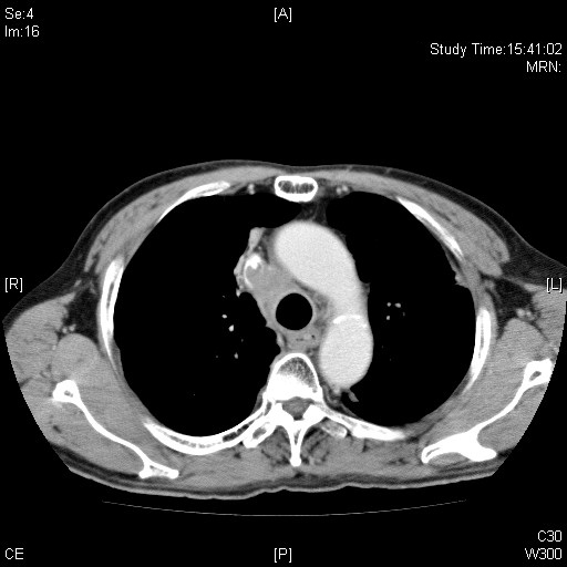
CT thorax taken few months before the current ICU admission showing marked eccentric compression on the adjacent confluence of the central veins including the superior vena cava.
A patient was admitted to our ICU for close monitoring after regional thrombolysis of thrombus in left brachiocephalic vein and superior vena cava. In reviewing his past history, there were images of recurrent stenosis and thrombosis of his major venous system that I would like to share with you. Also, various techniques for recanalization have been adopted.
He has known:
1. Chronic obstructive pulmonary disease
2. Stage IIIB non-small cell (large cell) cell lung carcinoma
– SVC and tracheal compression, tracheobronchial stent done but dislodged twice
– Thoracic RT (60Gy/30fr) with concurrent chemo completed 2 years ago in 12/2007
– disease progression with SVCO in 11/08
To revise the gross anatomy of the neck and thoracic venous system, see the website of The Department of Anaesthesiology of the University of Hong Kong here.
The initial CT thorax is shown below: 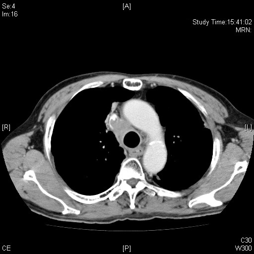
CT thorax showing marked eccentric compression on the adjacent confluence of the central veins including the superior vena cava.
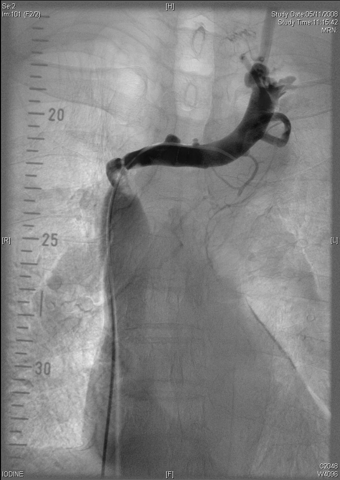
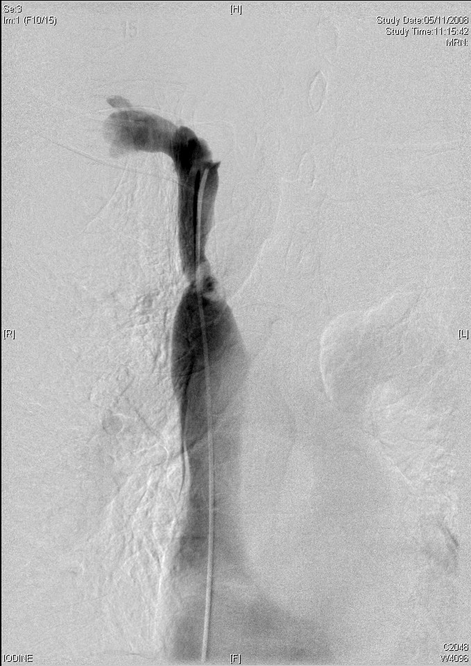
A venogram was done. Smooth marked eccentric stenosis is noted at the central vein confluence suggestive of extrinsic compression. A Wallstent (18mm x 60mm) was deployed across the left brachiocephalic vein and SVC crossing the stenosis. The stent was further dilated using 14mm balloon. Post-stenting angiography showed good angiographic result.
 |
 |
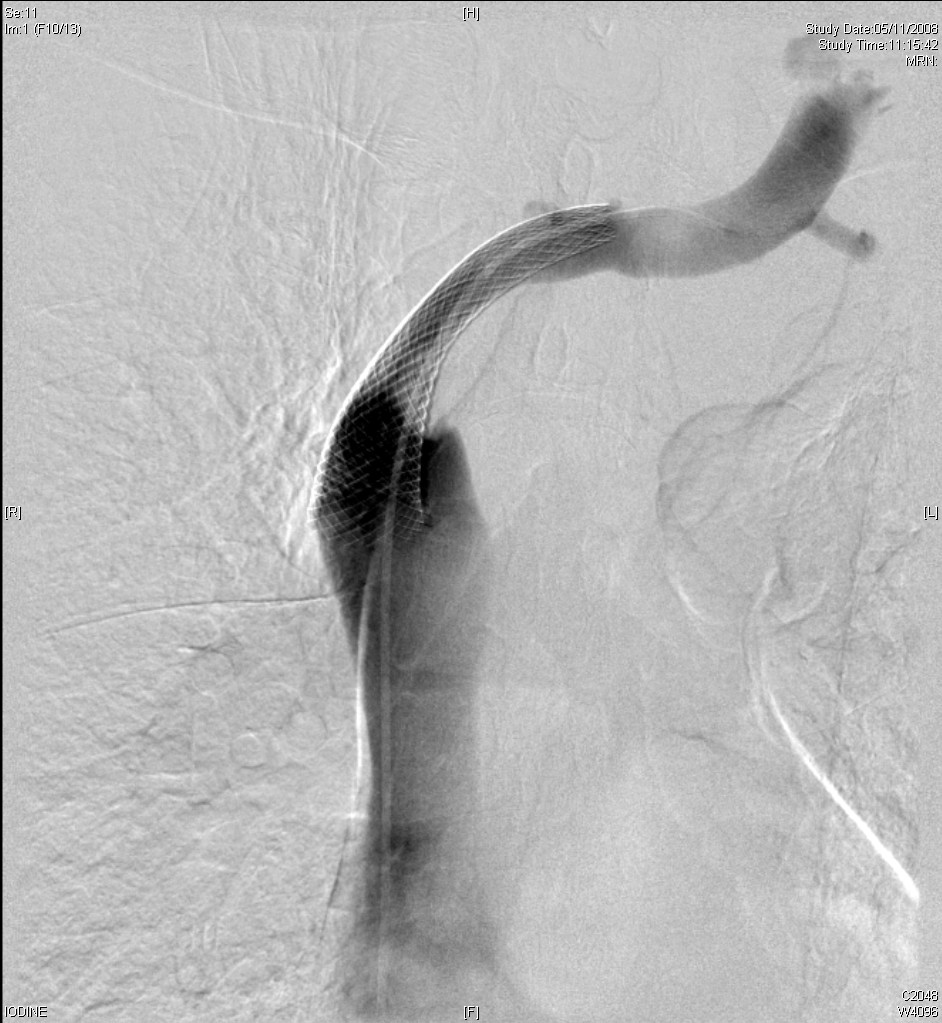 |
| Pre-stenting: L brachiocephalic vein and superior vena cava, extrinsic compression at the confluence is seen | Pre-stenting: R Brachiocephalic vein and superior vena cava, extrinsic compression at the confluence is seen | Post-stenting: L Brachiocephalic vein and superior vena cava |
One month after the above procedure, he complained of increasing shortness of breath with periorbital oedema. P/E showed engorged neck veins and UL swelling. A CT thorax was done: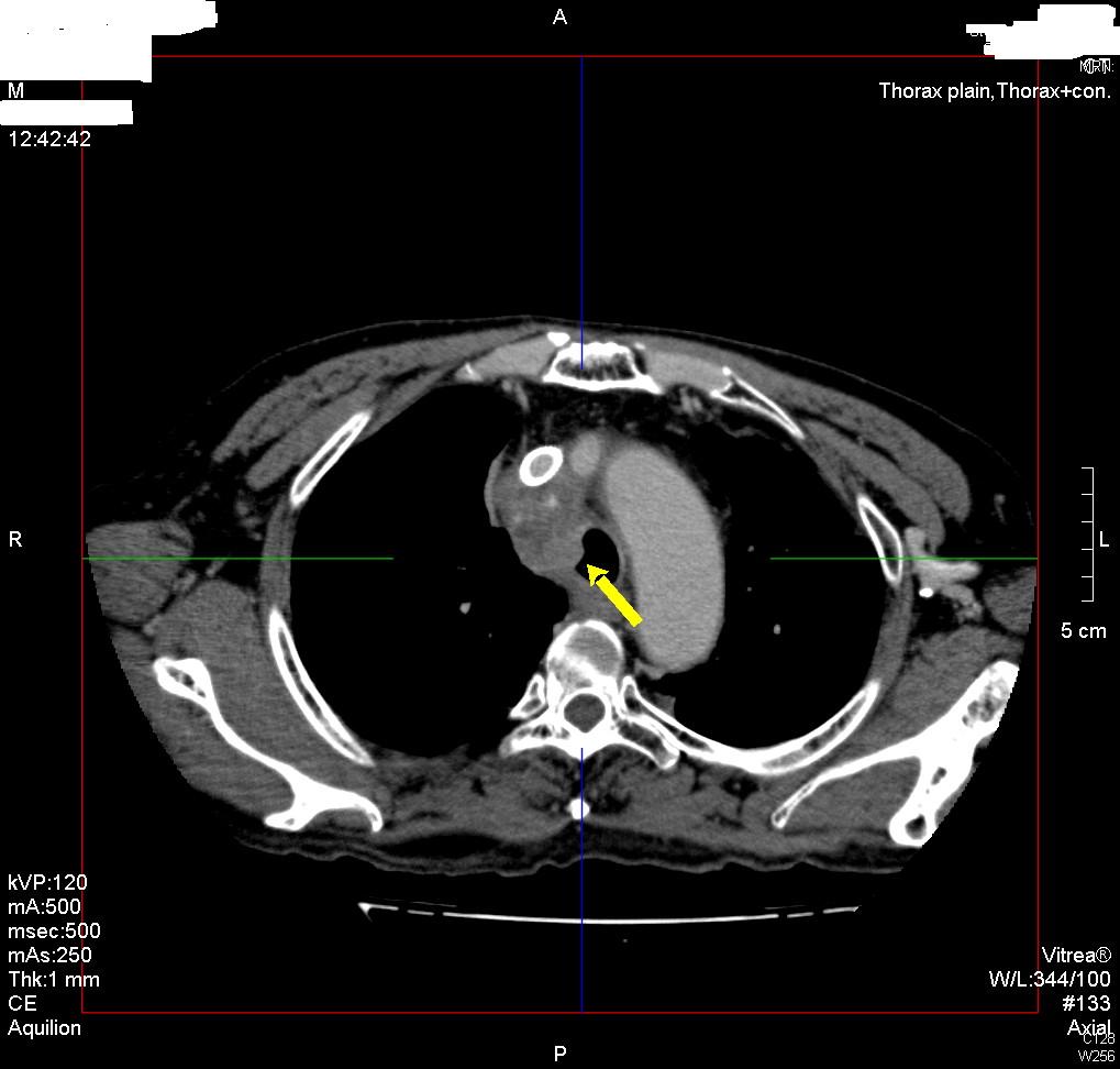
CT Thorax showed that the SVC stent from the left brachiocephalic vein down to SVC was patent with just small amount of eccentric filling defects, but the right internal jugular and right brachiocephalic vein were NOT opacified. The mediastinal mass lesion (yellow arrow) with intralesional calcified foci up to 40.3mmx32.6mmx45.8mm (APxTSxLS) is noted and indents right side of trachea.
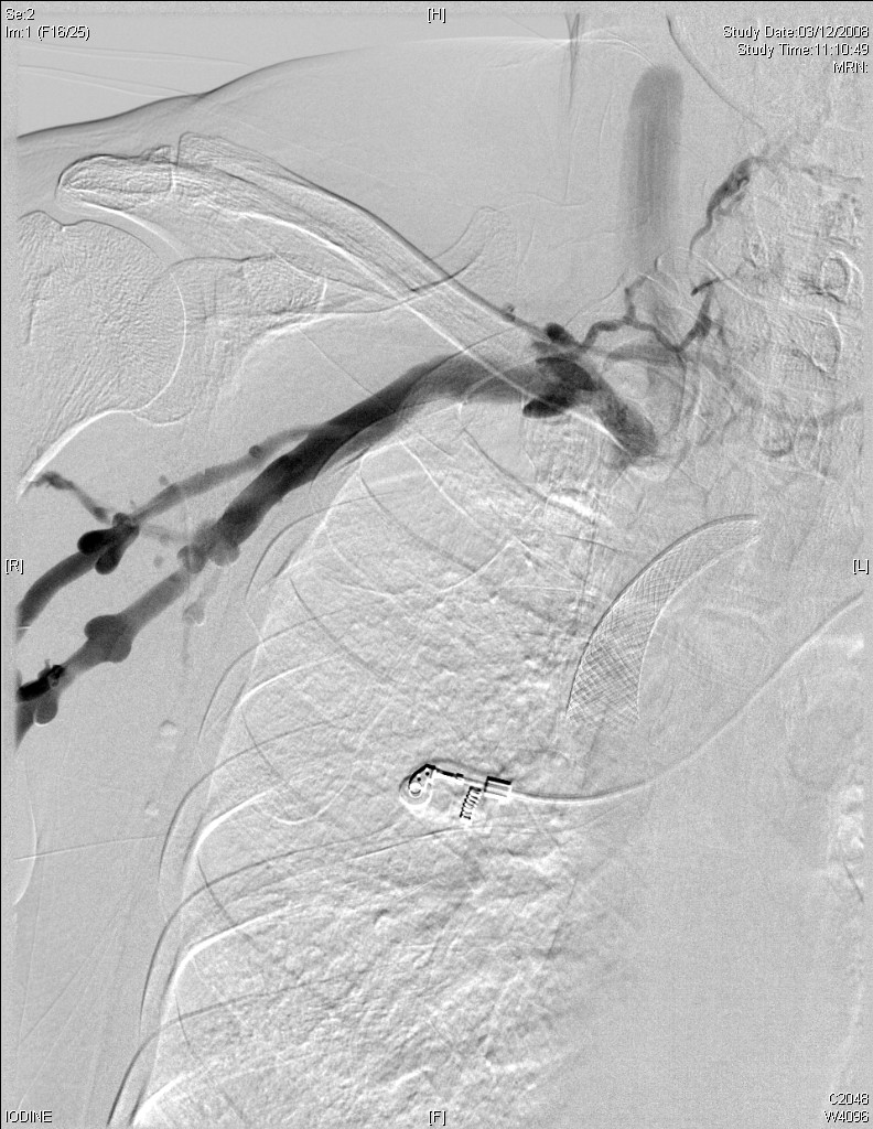
A RIGHT UPPER LIMB AND SVC VENOGRAM was done:
 |
 |
 |
|
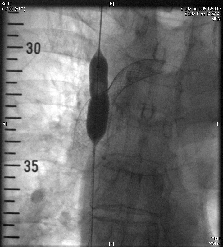
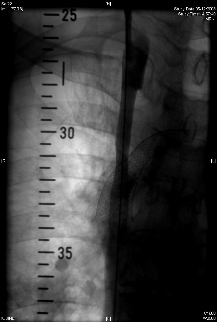
| RIGHT UPPER LIMB AND SVC VENOGRAM: The right basilic vein and right internal jugular vein are patent with very slow contrast flow. Marked narrowing of the right brachiocephalic vein is noted, likely due to invasion or extrinsic compression by tumour rather than thrombus. | Smooth marked eccentric stenosis is noted at the right brachiocephalic vein suggestive of extrinsic compression. The left brachiocephalic-SVC stent was noted in-situ. The wiremesh of the indwelling stent was traversed and dilated using 12mm balloon. | A Wallstent (18mm x 60mm) was deployed across the right brachiocephalic vein stenosis. The stent was further dilated using 12mm balloon (not shown). Post-stenting angiography showed good angiographic result. |
He was pllanned for anticoagulation therapy to prevent venous thrombosis. Allimta was given for 2 cycles.
Allimta is Pemetrexed, information is available here. For lung cancer treatment, there are two indications:
1. Initial Diagnosis of Advanced or Metastatic Nonsquamous Non-Small Cell Lung Cancer
ALIMTA (pemetrexed for injection) is approved by the FDA in combination with cisplatin (another chemotherapy drug) for the initial treatment of advanced nonsquamous non-small cell lung cancer (NSCLC), a specific type of NSCLC. ALIMTA is not indicated for patients who have a different type of NSCLC called squamous cell.
2. If Advanced or Metastatic Nonsquamous Non-Small Cell Lung Cancer Returns (our patient)
ALIMTA is approved by the FDA as a single agent (used alone) for the treatment of patients with advanced nonsquamous non-small cell lung cancer (NSCLC), a specific type of NSCLC, after prior chemotherapy. ALIMTA is not indicated for patients who have a different type of NSCLC called squamous cell.
A CXR was taken afterwards:
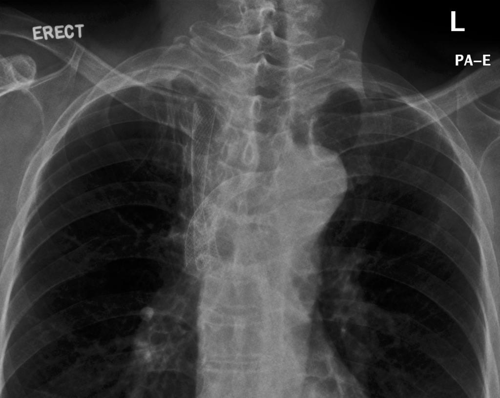
CXR showing the two stents-in-situ
About 2 months later, the patient was admitted for stridor, facial puffincess, bilateral upper limb swelling and fresh hemoptysis. Warfarin was stopped. CXR ↑RUL mass. A CT Thorax was done:
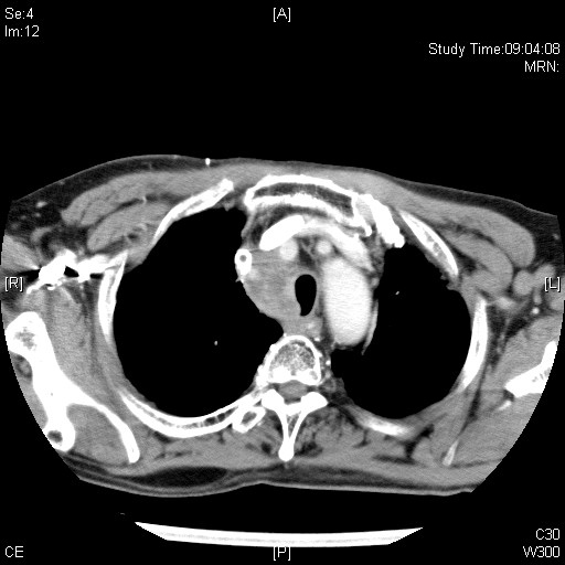
Patent left SVC stent with eccentric filling defect. Right SVC stent shows no contrast opacification, may represent thrombosis. The previously noted mediastinal mass lesion with intralesional calcified foci shows mild interval increase in size. Interval increase in indentation on right side of trachea is noted. Irregularity is noted at the right side of trachea, ?invasion.
Thrombolysis and angioplasty to SVC were done:
 |
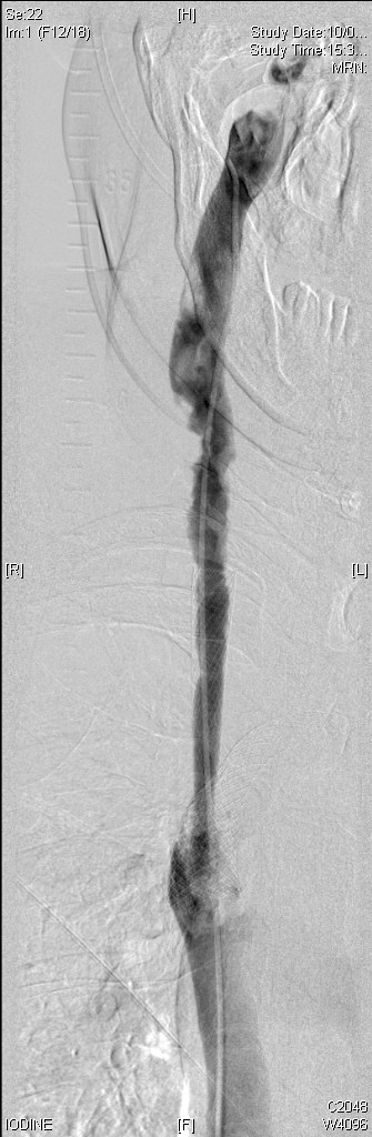 |
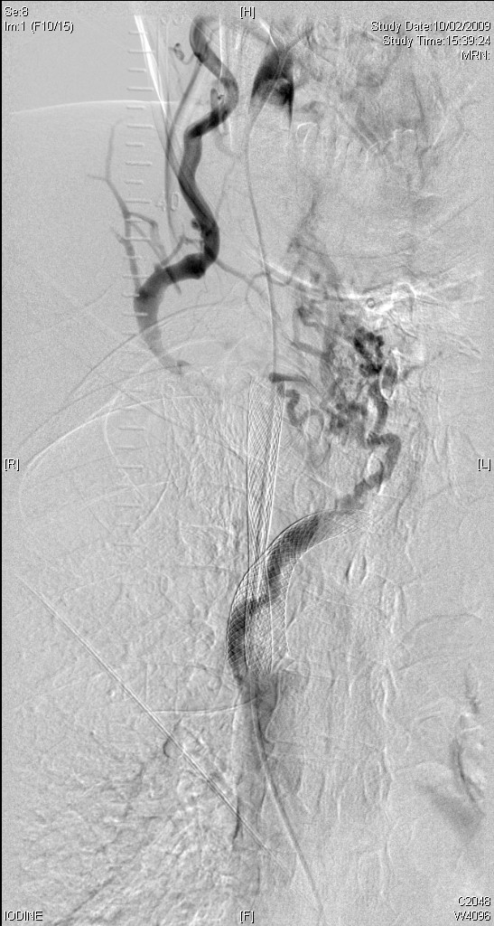
| Pre-thrombolysis and angioplasty:The thrombosed superior vena cava, the right brachiocephalic vein and the right internal jugular vein are successfully traversed via right common femoral approach using 7Fr vascular sheath and 5Fr H1 catheter. The superior vena caval stents are noted in-situ. Multiple intraluminal filling defects are noted in the right internal jugular vein, the right brachiocephalic vein and the superior vena cava; with numerous collaterals going to left brachiocephalic vein. The superior vena caval stents are noted in-situ. Multiple intraluminal filling defects are noted in the right internal jugular vein, the right brachiocephalic vein and the superior vena cava; with numerous collaterals going to left brachiocephalic vein. | Post throbolysis and angioplasty: rt-PA is given. Angioplasty of the right superior vena cava stent and maceration of the thrombi is done using 12mm and 14mm balloons and Fogarty balloon. Then, Post-thrombolysis and post-angioplasty venogram show few small non-occlusive residual thrombi in the right internal jugular vein and superior vena cava. There is good flow of contrast via the right superior vena cava stent to the right atrium. |
A month later, the patient was readmitted for bilateral UL and facial swelling and SOB.
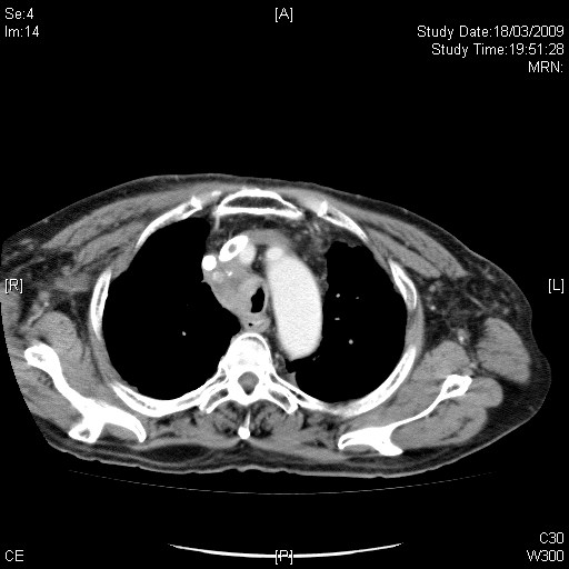 |
CT thorax with contrast:
IMPRESSIONS: |
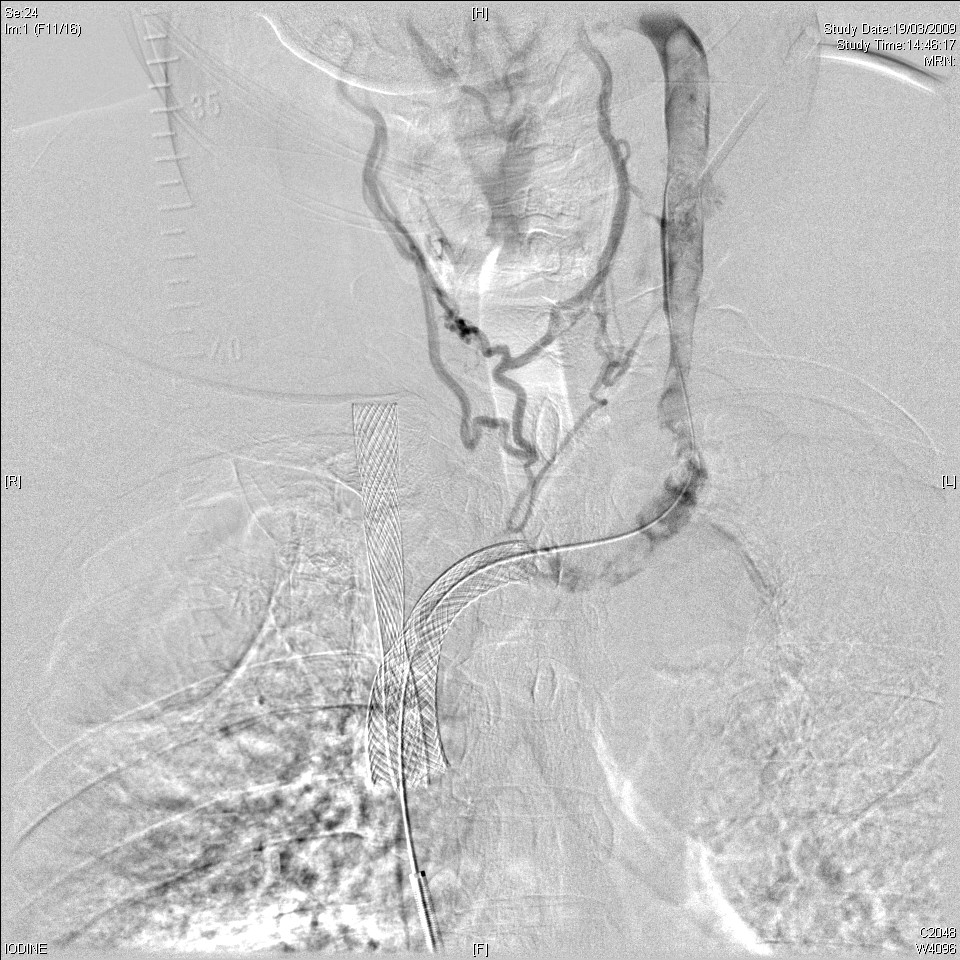
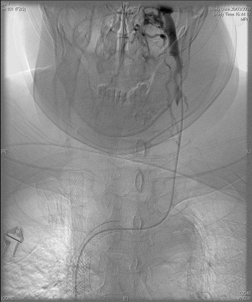
SUPERIOR VENA CAVOGRAM & THROMBOLYSIS & THROMBECTOMY OF THE THROMBUS IN THE LEFT INTERNAL JUGULAR VEIN, LEFT BRACHIOCEPHALIC VEIN-SVC STENT were attempted in daytime. The patient was admitted to ICU for close montoring after the procedure.
 |
 |
 |
|
The venograms were performed via right femoral approach using 7 French vascular sheath and left internal jugular approach using 5 French vascular sheath. The right internal jugular vein, the right brachiocephalic stent are patent with few small non-occlusive intraluminal filling defects suggestive of thrombi. Left brachiocephalic and superior vena cava (SVC) venogram shows long segment of thrombus extending from the left internal jugular vein (up to level of angle of mandible) to the left brachiocephalic/SVC stent. |
The thrombi in the left internal jugular vein and the left brachiocephalic/SVC stent are macerated using 5mm balloon and Fogarty balloon. The left brachiocephalic/SVC stent is dilated using 12mm balloon. rtPA thrombolysis using pulsed spray technique is performed.
|
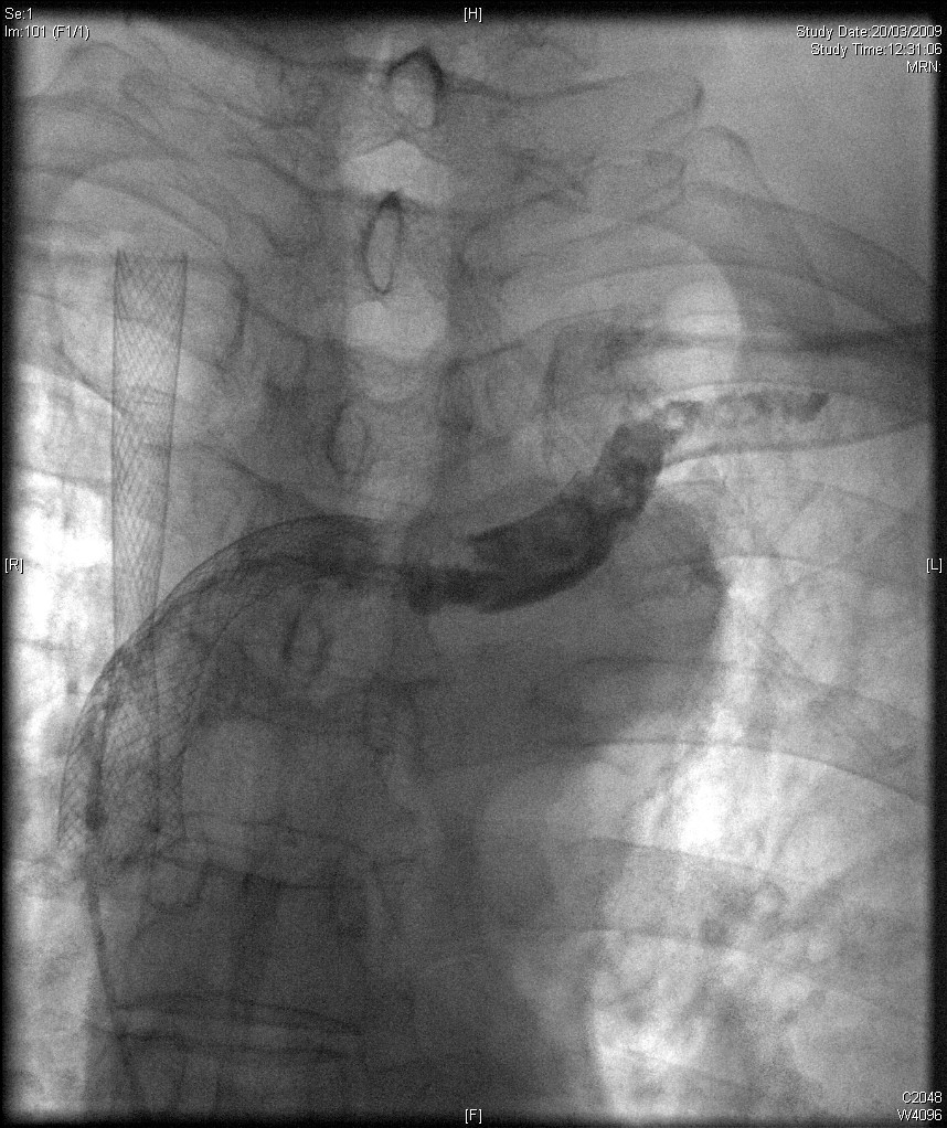
Post-procedural left brachiocephalic-SVC venogram demonstrates thrombi in the left internal jugular vein and the left brachiocephalic/SVC stent. In view of the risk of hemorrhage, further thrombolysis is not performed. Technically unsuccessful thrombolysis of the left internal jugular vein and the left brachiocephalic/SVC stent. |
Because of the failed thrombolyis, embolectomy and angioplasty were performed few hours afterwards with 6F Fogarty catheter, 6mm, 8mm and 10mm balloon. Totally, 3000 heparin IV was given. Post-procedure venogram (below) revealed marked decrease in amount of intraluminal filling defects with few non-occlusive thrombi in the left jugular vein and in the left SVC stent. In view of risk of hemoptysis, further thrombolysis was not performed.
|
{morfeo 25} |
He was started on heparin. Patient’s facial edema gradually improved again and was discharged from ICU uneventfully.