
A CXR taken 2 years ago is shown in figure 2.
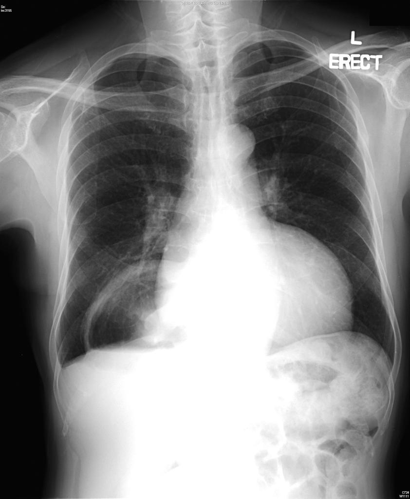
Figure 2
Diagnosis: Right hiatus hernia
In Figure 1, the NG tube was actually in the stomach. Because of worry about malpositioning, the NG tube was removed and reinserted, but another CXR gave similar finding. Old film (Figure 2) was then compared. Correct positioning of the NG tube was eventually confirmed by pH testing as well as direct DL visualization.