
Submitted by Dr Ho Chun Ming Chris on 14 April 2009
ICU, North District Hospital
A 58 year-old ex-smoker presented to the Accident and Emergency Department because of coffee ground vomiting. He was admitted to our ICU for hypovolemic shock and acute kidney failure. There was no abdominal pain, chest symptoms or fever.
Physical exam:
– BP 103/49, Sinus tachycardia with 120/min, SpO2 93% at RA, Afebrile, Clinically dehydrated
– Abd: soft, no signs of peritonism, no malena
– Other examination were unremarkable
Bld tests:
– severe leukocytosis, acute kideney failure and metabolic acidosis
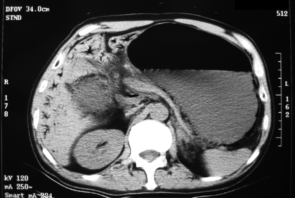
Figure. CT Abdomen
Click Read More to see the answer.
Answer: Emphysematous gastritis
The above CT showed that the abdomen was grossly distended. Interstitial gastric emphysema (gas in the stomach wall) was found. Duodenum and small bowel were not distended. There was no pneumatosis intestinalis. Gas was found in the portal vein. There was no ascites or peritoneal gas.
The patient underwent diagnostic laparoscopy and urgent OGD in the operating theater.
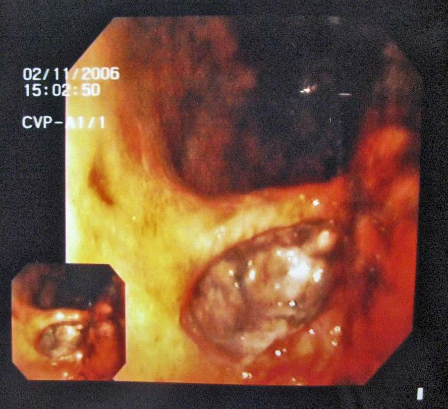
Figure. Image of OGD
OGD showed huge cavitating chronic gastric ulcer obstructing pylorus
Diagnostic laparoscopy: viable stomach wall, clean peritoneal cavity without perforation or ischaemia, peritoneal fluid was clean
Culture of peritoneal fluid: negative.
We planned for gastrectomy if patient deteriorated. Our patient survived with conservative treatment with ampicillin, cefuroxime, and metronidazole. There was also full recovery of renal function after a short course of CRRT.
Discussion
Emphysematous Gastritis
Incidence: A rare condition, 41 cases reported from 1889-2000
Background: The stomach is normally resistant to invasion because of excellent blood supply, acid pH, and mucosal barrier. Air within the stomach wall is usually produced by gas-forming organisms. This condition was first described as a clinicopathological entity by Fraenkel in 1889. (A 35-year-old man having debridement for a crush injury and subsequently died after several episodes of hematemesis, abdominal pain, and diarrhea). As a radiological diagnosis it was first described by Weens in 1946.
Radiology: It was characterized by intramural gas within a thick-walled, dilated stomach, gas in portal venous system. It appears within the first 1-2 day of the illness and can persist up to a month.
Differential diagnosis: gastric emphysema, and phlegmonous gastritis
Most common predisposing factors: ingestion of corrosive substances, alcohol abuse, abdominal surgery, diabetes mellitus, immunosuppression
Most commonly involved organisms: Streptococci, Escherichia coli, Enterobacter species, Clostridium welchii, and Staphylococcus aureus
Diagnosis: Computed tomography (CT) is the diagnostic procedure of choice
Mortality rate: 60-80%, gastric contractures after recovery were noted in 10%
Management: antibiotics covering gram-negative organisms and anaerobes, and surgery in appropriate cases may enhance survival