
Submitted by Dr LAU Chun Wing Arthur on 25 Feb 2009; Department of Intensive Care, PYNEH
M/34, social smoker + drinker; PHX: Diabetes mellitus, initially on insulin, later switched to oral hypoglycemic agents. Defaulted FU, on over-the-counter diabetic medications
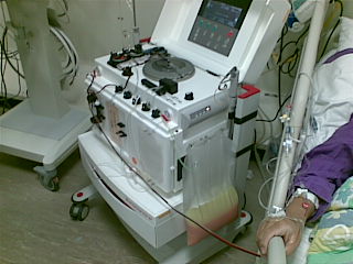
Admitted to another hosptial for epigastric pain for 3 weeks, transferred to us for further care. We regarded that plasmapheresis was necessary for the treatment.
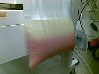
Plasma of patient obtained by the centrifugation method of plasmapheresis
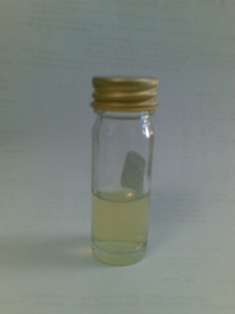
Plasma of another similar patient by the plasma filtration method of plasmapheresis
What is their common diagnosis?
Click here to read more.
Dx: hypertriglyceridemia-induced or related acute pancreatitis
This patient has had acute pancreatitis before. This time, the patient’s amylase was 1064 with normal LFT. Triglyceride level was 68 (grossly elevated) before transfer, glucose control was poor. US and CT confirmed diagnosis of acute pancreatitis. TG level decreased to 12 on fasting alone. In view of the still high TG level, plasmaphoresis was started. The pictures above show chylomicron-rich plasma removed from the patients.
TG level remained static at around 4 afterwards and epigastric pain subsided. He was seen by endocrinologist who suggested to continue insulin at the moment, and may add lopid later.
CT repeated in our hospital:
{morfeo 15}
Stranding is noted around the pancreas. Pancreas is prominent in size. No parenchymal calcification is noted. Parenchymal enhancement of the pancreas is noted. Heterogenous enhancement with areas of fat density is noted at the uncinate process, may represent fatty infiltration or edema. A 1.3cm hypodense lesion is noted at the neck of the pancreas. Wall of duodenum appears thickened. Distal splenic vein, distal inferior mesenteric vein and origin of main portal vein appears narrowed, ?related to compressive effect of edema. CT features are compatible with acute pancreatitis. No definite CT evidence of pancreatic necrosis.
Discussion
Athyros et al 2002: Long-term outcome study
Berger et al 2001: Insulin and heparin may also help reduce the TG level
Kimura W et al 1996: Rat study showing that pancreatic damage initiated via different pathogenetic pathways can be increased by triglycerides in a dose-dependent fashion (may imply that TG level has to be decreased as much as possible as an important treatment during acute pancreatitis)
Furuya T 2002: Two hypertriglyceridemic acute necrotizing pancreatitis patients. One underwent plasma exchange and recovered, the other did not and died.
Fortson et al 1995: This study tried to classify hyperlipidemic acute pancreatitis into three common clinical presentations: 1. The most common presentation is a poorly controlled diabetic with a history of hypertriglyceridemia. 2. The second presentation is the alcoholic found to have hypertriglyceridemia or lactescent serum on admission. 3. The third, about 15-20% of patients, is the nondiabetic, nonalcoholic, nonobese patient with drug-or diet-induced hypertriglyceridemia.