
Submitted by Dr LAU Chun Wing Arthur on 7 March 2009
Department of Intensive Care, Pamela Youde Nethersole Eastern Hospital
This F/79 patient had past medical history of hypertension, hyperlipidaemia, mitral and tricuspid regurgitation, history of non-ST elevation MI, congestive heart failure, peripheral vascular disease, bilateral renal artery stenosis. In recent two months she was admitted for passing altered blood. Haemoglobin level was 7 g/dL. Upper endoscopy, colonoscopy and capsule endoscopy could not identify the exact source of bleeing, and was therefore discharged and planned for further workup. This time she was admitted for expectoration of blood. The CXR is shown below:

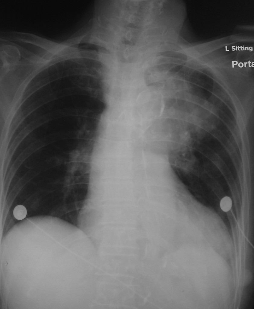
What is the diagnosis? Click “Read More” for the answer.
Urgent OGD: blood seen around vocal cords with fresh blood and clot coming down from esophagus, total 600ml blood suctioned. Post-OGD BP lowish with mild tachycardia, SpO2 95% on 4L/min O2 via nasal cannula.
She was transferred to the ICU for further management.
An urgent CT thorax and angiogram of the aorta was done:
| {morfeo 17} |
There is a focal irregular dilatation in the proximal descending aorta distal to the origin of the left subclavian artery, measuring about 2.8cm x 3.0cm x 3.0cm, suggestive of focal aneurysmal dilatation. Thickened eccentric hyperdensities measuring up to 2.4cm in thickness is seen in the left lateral aspect of the dilatation, which displaces the intimal calcification of the proximal descending aorta laterally. Features are worrisome of a mural thrombus/hematoma and aneurysm/pseudoaneurysm formation involving the proximal descending aorta. Atherosclerotic changes with wall calcifications are seen along the aorta. The external caliber of the descending aorta and abdominal aorta are within normal upper limits with no definite focal aneurysmal dilatation seen. No evidence of intimal flap is seen. The main innominate, left common carotid, left subclavian, celiac trunk, superior mesenteric artery and bilateral renal arteries are opacified. Ground glass opacity/consolidative changes are seen diffusely over the left lung.
Discussion
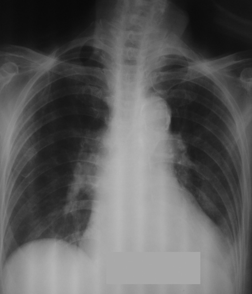
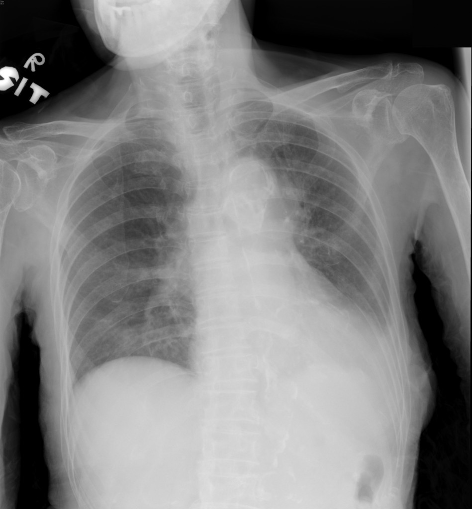
Old CXRs were reviewed:
 |
 |
|
From left to right: 2 months before admission (calcifications at aortic arch seen); 12 days before admission (some bulging of proximal descending aorta with displacement of calcifications, left lower lobe infiltrates); this admission (gross bulging of aorta with surrounding lung infiltrates due to blood)
This patient had a rapidly expanding and leaking aortic aneurysm which had probably eroded into the esophagus as assessed on the CT thorax, and she developed “occult” GI bleed and eventually massive bleeding and aspiration. This patient had all the markers of underlying atherosclerosis from the history of her past health, and because of the poor premorbid state, she declined further invasive treatment.