
Submitted by Dr Anne KH Leung on 6 April 2009
Queen Elizabeth Hospital, Hong Kong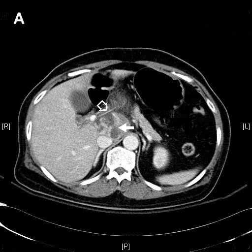
Figure 1. CT abdomen
A 55-year-old woman presented with one week history of abdominal pain, fever, chills and rigor. Initial blood tests showed leucocytosis (WCC = 29.8×10^9), renal impairment and mildly elevated alkaline phosphatase on liver function test. She was admitted to the Intensive Care Unit for septic shock. Blood culture grew Streptococcus milleri. Urgent CT abdomen (Figure 1) was done and is shown above.
The CT thorax reviewed a large lobulated and cystic mass in the caudate lobe of the liver suggestive of liver abscess (White solid arrow). There was also a thin linear hyperdensity (White hollow arrow) lying horizontally in close proximity to the portal vein, suggestive of a foreign body but of unknown nature. There was no free gas in the abscess or around the foreign body that could suggest visceral perforation. Emergency laparotomy was performed and revealed a 4 cm slender fish bone-like foreign body (Figure 2). The abscess cavity inside the caudate lobe was drained. Also, a 3 cm induration was noted at the lesser curve of the stomach wall and was sealed with omentum, and this was highly suggestive of previous perforation by some foreign body. The patient made a rapid recovery after the operation and was discharged home 8 days afterwards.

Figure 2