
Submitted by Dr LAU Chun Wing Arthur on 19 Feb 2009
Department of Intensive Care, Pamela Youde Nethersole Eastern Hospital
F/37, c/o Massive hemoptysis while at work.
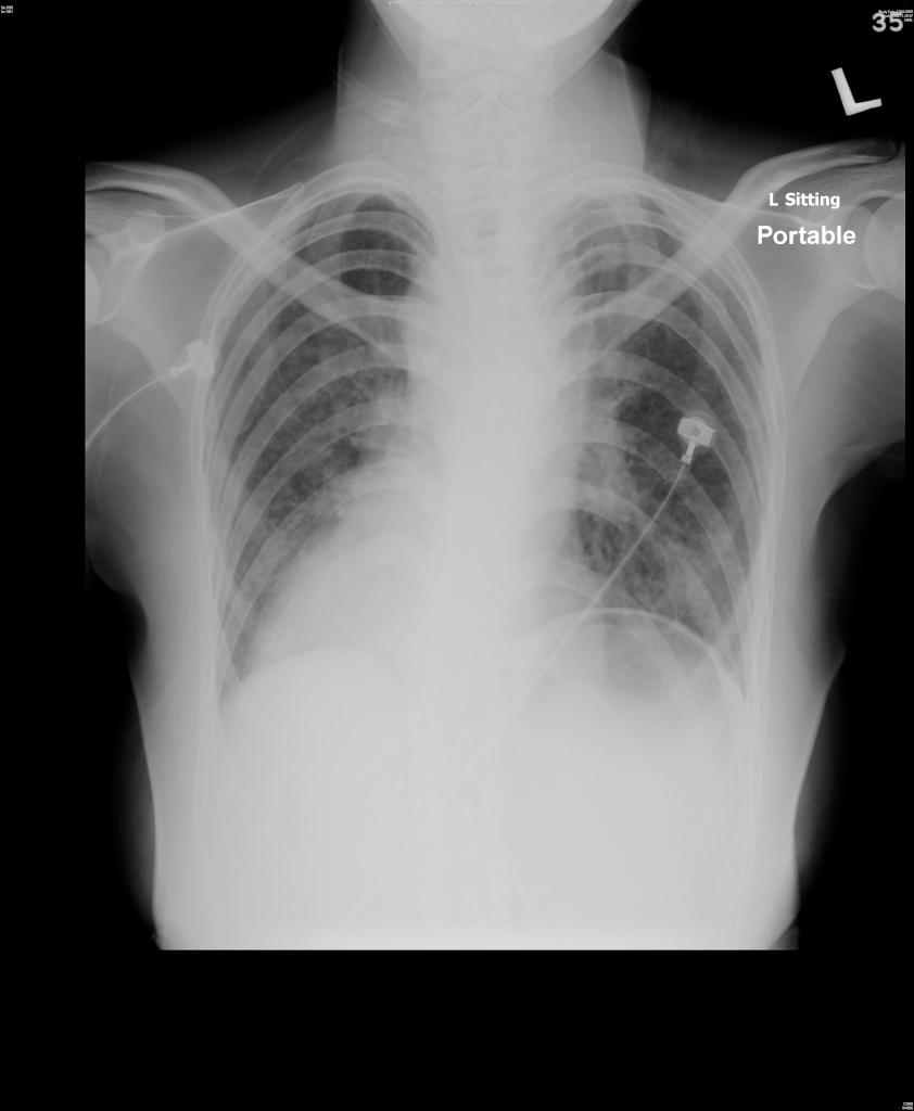
CXR (click image to enlarge)
Comment on this CXR. What are the abnormalities and where is the site of bleeding?
Click here to read more.
This patient actually had the following medical history:
1. Complex cyanotic heart disease: situs solitus/dextrocardia, double inlet right ventricle (DIRV), double outlet right ventricle (DORV), common AV valve with mild to moderate regurgitation, PS, RPA stenosis. Left classical BT shunt in more than 26 years ago, not for Fontan operation due to high OT risk
2. Polycythemia requiring repeated venesection
3. Hx of acute cerebral infarction in 2 years ago
4. Recurrent hemoptysis: CT thorax 5 years ago showed no lung mass
Bronchial arterial embolisation was done.
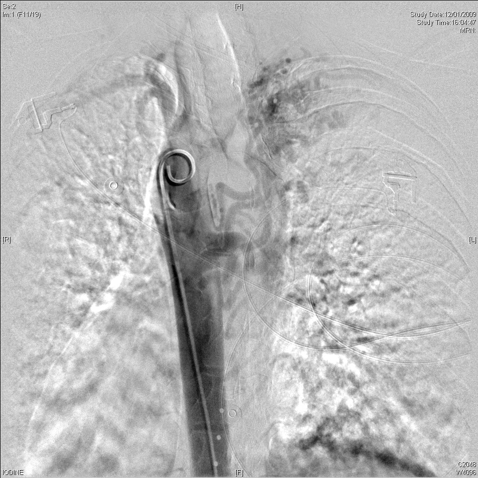
Aortogram
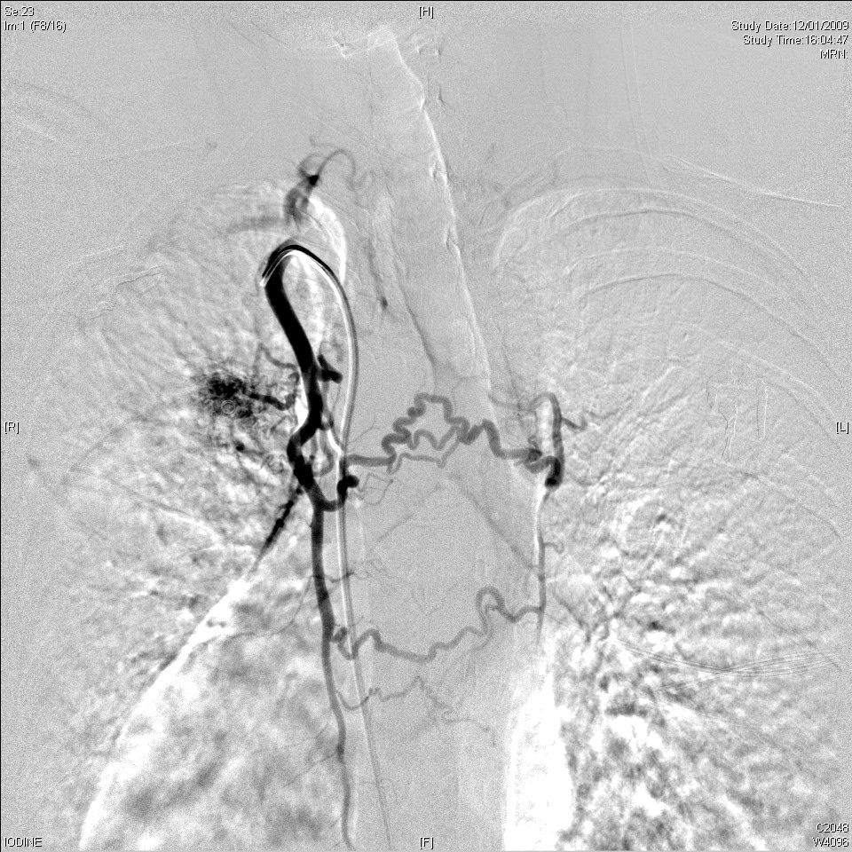
R internal mammary arteriogram
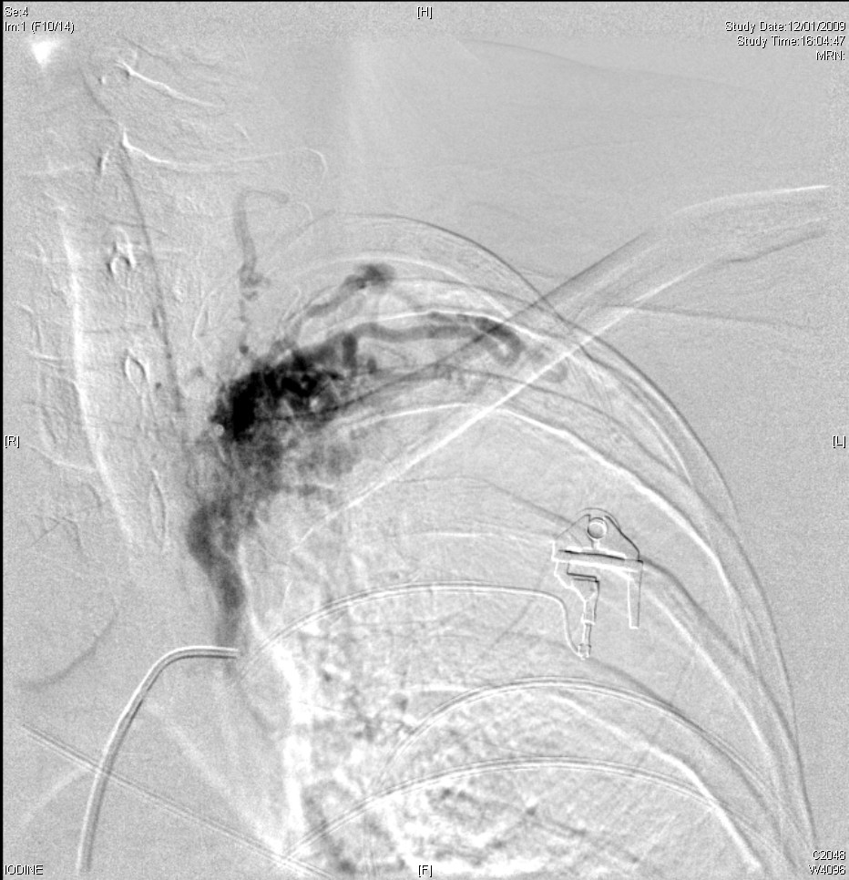
L 3rd intercostal arteriogram
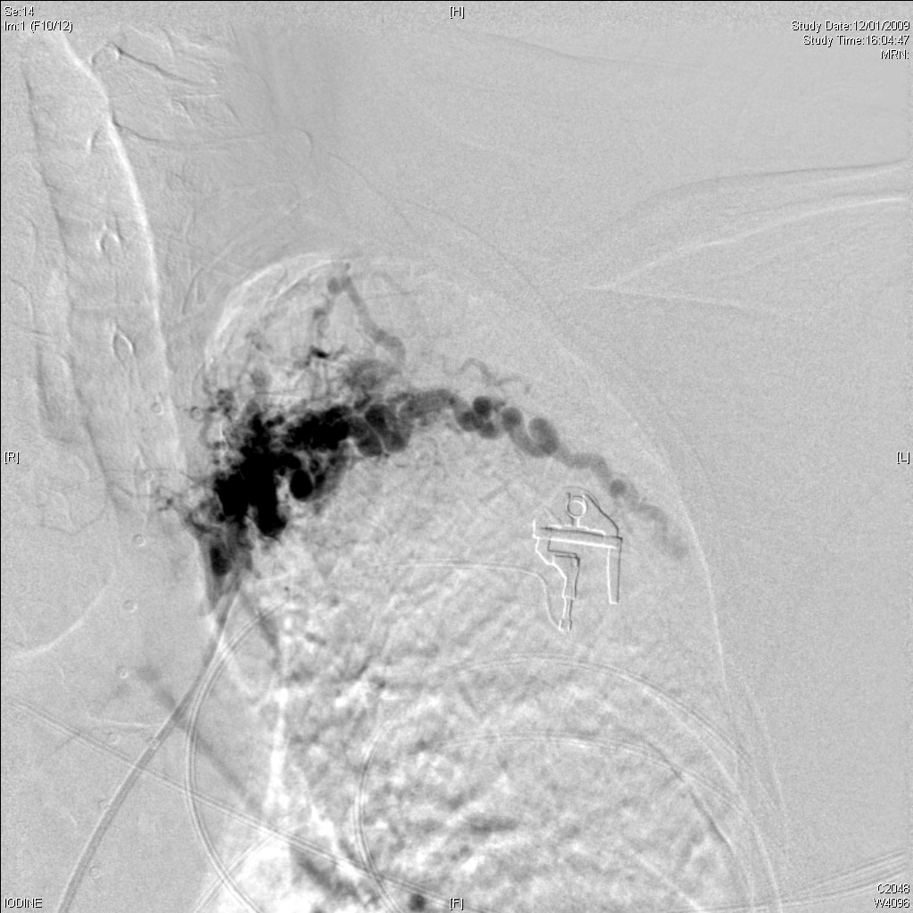
L 4th intercostal
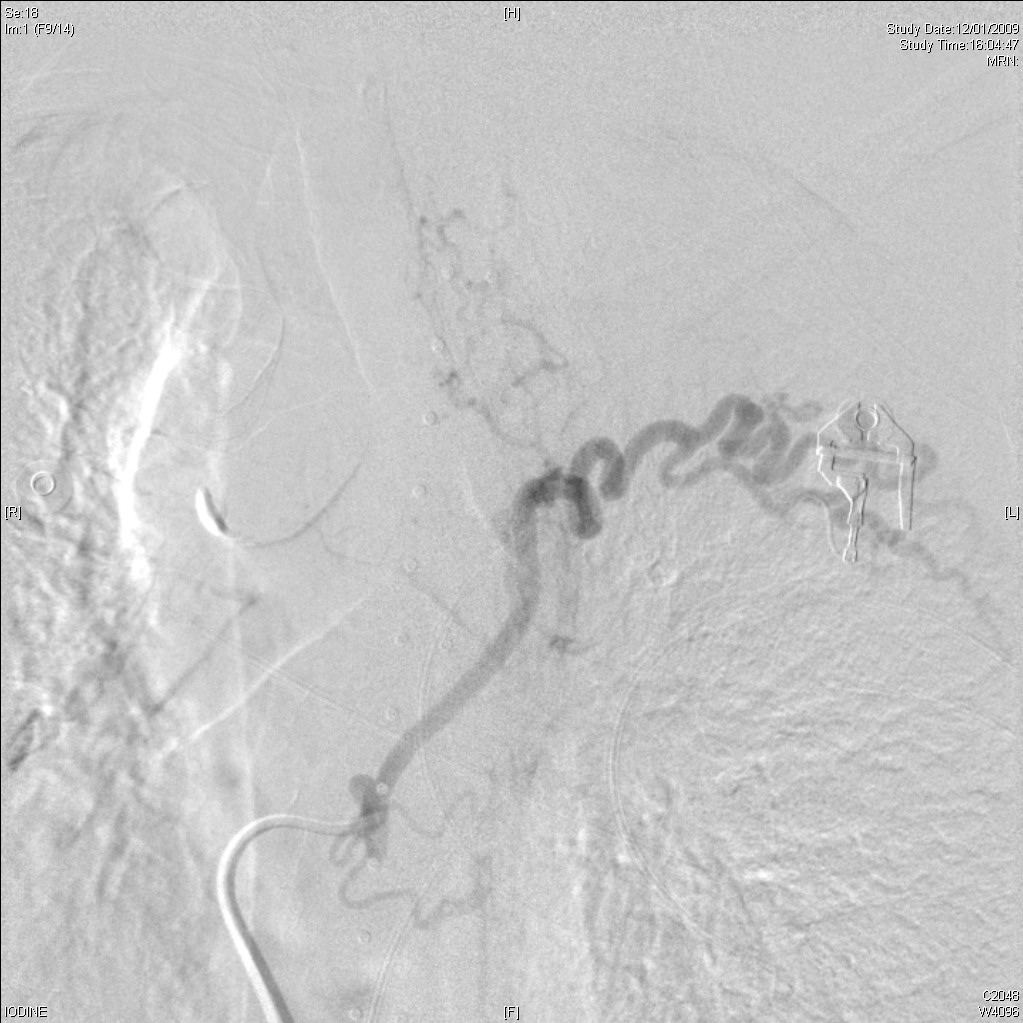
L 5th intercostal
What are the abnormalities you can pick up so far?
The CXR shows dextrocardia and bilateral lower lung infiltrates due to aspirated blood, especially on left side. The view is quite anti-lordotic, and the upper few ribs overlap each other. However, we can still see rib notching over the inferior portions of left 3rd to 5th ribs and even the 6 ribs.
Bronchial arteriogram with embolisation (BAE) was performed. We can already vaguely see the marked hypertrophy and tortuosity of the left intercostal arteries with marked peribronchial hypervascularity and bronchio-pulmonary communication in the aortogram, and certainly more obviously in individual intercostal arteriograms. Selcetive cannulation of the hypertrophied left intercostal branches supplying the left upper lobe using 5Fr Cobra 1 and Shephard hook catheters. These was followed by embolization using 710-1000 micron Ivalon and gelfoam. No dilated R bronchial artery is detected. The R internal mammary artery was also found to be hypertrophied with branches supplying the right upper lobe. Selective catheterization and embolization was performed using 5Fr C1 Cobra catheter and gelfoam. The procedure was well tolerated with no immediate complications. The exact cause of the multiple tortuous intercostal arteries is not certain.
Learning point
Look for rib notching over the inferior portion to suspect the presense of hypertrophied and tortuous intercostal arteries