
Submitted by Dr Lily LL Chang, ICU, PYNEH, Hong Kong, on 25 Aug 2009
A 25 year-old lady with history of SLE with nephritis, complicated with ESRF on CAPD had been admitted one year ago for headache, vomiting, impaired conscious state and convulsion. MRI brain (T2/PD/Flair) showed the following lesion (Figure 1).
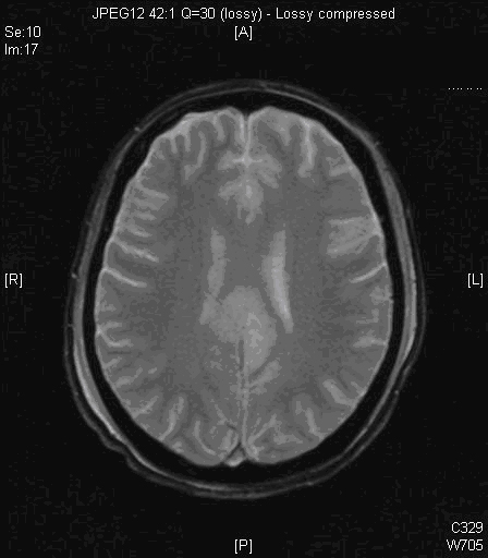
Figure 1
The lesion was completely reversible a month afterward (Figure 2).
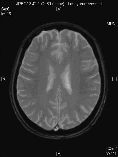
Figure 2
The lesion was compatible with posterior reversible encephalopathy syndrome (PRES) by MRI. MRI brain showed T2/PD/Flair hyperintense gyriform signal change over bilateral parasagittal region involving both grey and white matter (Fig 1). Subsequent MRI brain showed resolution of the confluent hyperintense gyriform signal change previously seen in the bilateral parasagittal region involving both grey and white matter (Fig 2).
A year afterwards, she was presented to us again with headache, vomiting and impaired conscious state. She was afebrile and without neck rigidity. She then developed generalized tonic-clonic seizure in AED and was aborted by diazepam. CT brain showed symmetrical hypodensities over bilateral thalamus, internal capsule, caudate nucleus and brainstem. She was intubated for airway protection due to poor conscious state.
She was accessed by neurologist and suggested the possible diagnosis of PRES again. The precipitatin factors this time were suspected to be poor compliance to dialysis and uncontrolled hypertension. MRI brain was done which showed extensive T2 and FLAIR patchy brightenings involving both supratentorial compartment and infratentorial compartment including the brainstem, pons and anterior columns of medulla; the findings are consistent with PRES (Fig 3). Blood pressure was controlled and dialysis regime was adjusted. Her conscious state had gradually improved and she was able to be extubated.
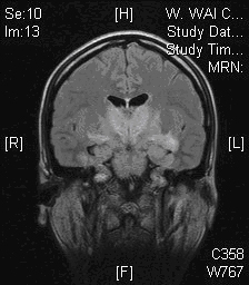
Figure 3
Discussion:
Posterior reversible encephalopathy syndrome (PRES), also known as reversible posterior leukoencephalopathy syndrome (RPLS), is a syndrome characterized by headache, visual loss, confusion and seizures. The possible etiologies include malignant hypertension, eclampsia, hypercalcaemia, and medications such as tacrolimus and cyclosporine.
The pathophysiology of PRES is under debate, but it is related to disordered cerebral autoregulation. Two pathophysiologic mechanisms have been proposed regarding cerebral autoregulation—cerebral vasospasm, which results in cytotoxic edema, and vasodilatation, which results in vasogenic edema. The latter is more favored by most experimental and clinical data. The pathophysiology of PRES also implicates endothelial dysfunction, especially in cases without severe hypertension, such as pre-eclampsia or cytotoxic therapies.
MRI is the most sensitive method for diagnosis. Typical MRI features include multiple cortoco-subcortical areas of hyerintense signal involving the occipital lobe, bilateral parietal lobes and the pons as shown in Fig 4. However, other structures (such as the brain stem, cerebellum, and frontal and temporal lobes) may also be involved, and although the abnormality primarily affects the subcortical white matter, the cortex and the basal ganglia may also be involved.
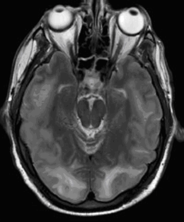
Figure 4
It is important to recognize this syndrome since it is completely reversible. Treatment includes correcting hypertension and electrolytes and lowering the dose of the involved drugs. Of course, other causes including CNS infection or cerebrovascular accident should be excluded.