
Submitted Dr OY Tam, ICU, PYNEH, Hong Kong on 9 October 2009
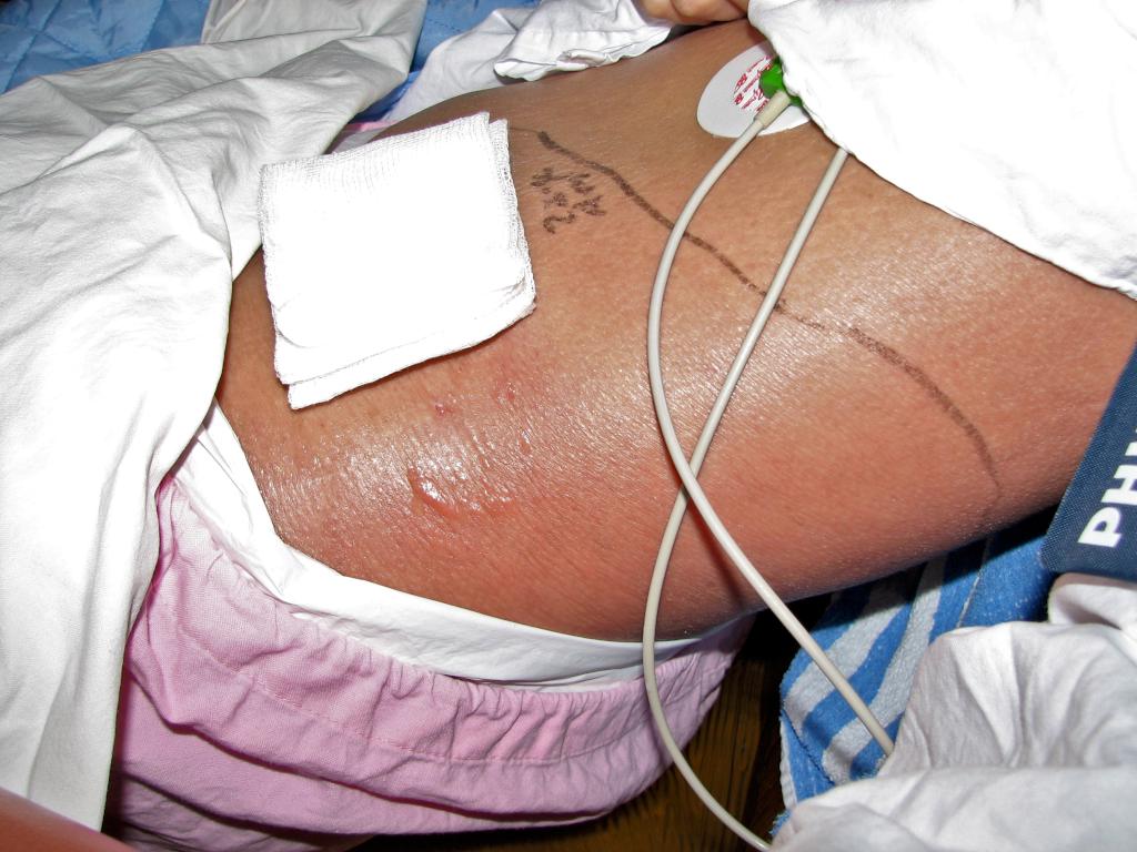
Figure 1. Picture showing the extent of erythema and blister formation. The gauze is covering the drained carbuncle wound.
A 30-year-old man with good past health was admitted to the surgical ward for high fever and a carbuncle with surrounding cellulits at left flank.
Haemodynamics on admission were stable. However, he developed septic shock within 24 hours of hospitalization. The erythema extended rapidly to involve the contralateral groin and the genitalia. He was transferred to ICU for further management. Blood tests showed disseminated intravascular coagulopathy, raised inflammatory markers and leukocytosis with left shift phenomenon. He was resuscitated with fluid challenge and required high dose vasopressor. Urgent CT did not show any sign of necrotizing faciitis. Ceftriaxone, doxycycline and metronidazole was empirically started. Yet, the extent of erythema progressed further with blister formation. (Fig. 1)
CT was repeated which showed increase in extent of subcutaneous soft tissue thickening and stranding involving the lower abdomen, pelvis, scrotum and thighs bilaterally. No gas pockets were seen. Still there was no radiological sign of necrotizing fasciitis. (Fig. 2)
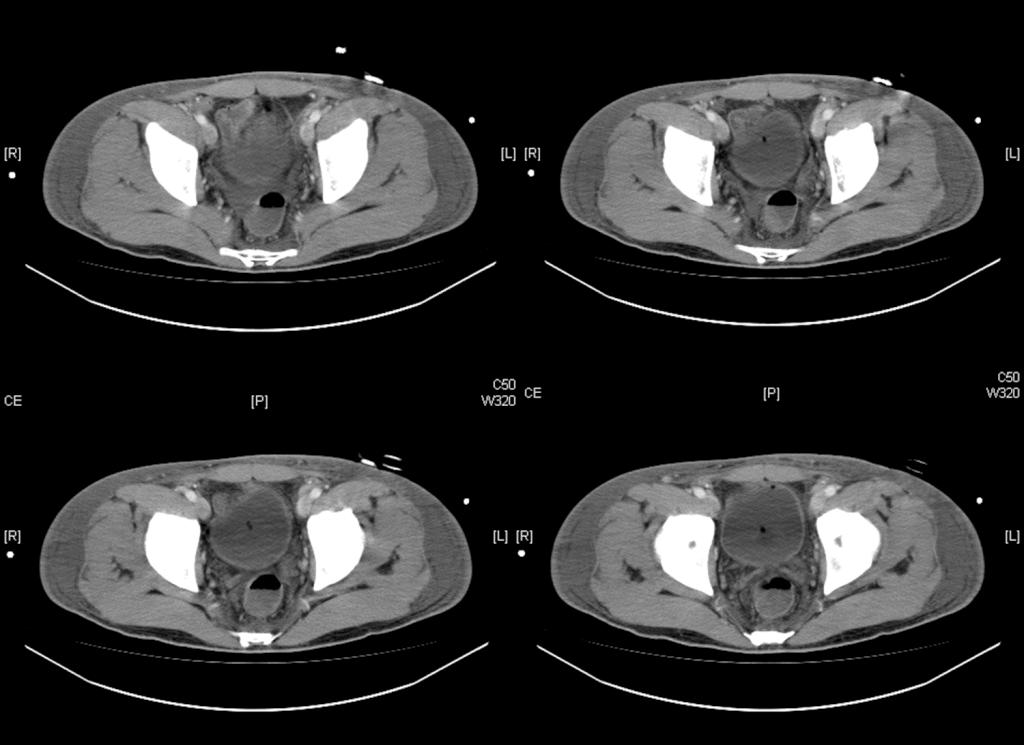
(Fig. 2)
Wound swab and tissue culture subsequently yielded Lancefield group A streptococcus pyogenes. Streptococcal toxic shock syndrome was suspected. Antibiotics were switched to clindamycin and ceftriaxone. IVIg was given daily for 5 days. He was gradually stabilized and discharged from the ICU.
In the general ward, the soft tissue infection persisted. Radical debridement and skin grafting was subsequently performed for him. During operation, marked induration was found over the left groin. All subcutaneous fat and fascia was necrotic with loculations of purulent discharge diffusely spaced over the indurated skin, supporting a diagnosis of necrotizing fasciitis.
Discussion
Necrotizing fasciitis (NF) is a life-threatening infection that affects the subcutaneous fat and deep fascia overlying muscles. Diagnosis is often missed because subcutaneous changes may not be readily apparent. Early diagnosis and prompt radical surgery decrease morbidity and mortality. The anatomy of skin is shown in the Figure 3.
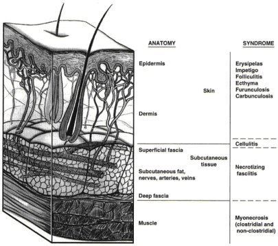
(Fig. 3)
Type 1 NF is a polymicrobial infection. It is often seen postoperatively or in patients with diabetes mellitus or peripheral vascular disease. Type 2 is caused by Streptococcus. Sometimes it is co-infected with Staphylococcus aureus. It can occur postoperatively or as a result of penetrating trauma, varicella infection, burns, or minor cuts. It is the most common form of NF in children and is characterized by rapidly extending necrosis and severe systemic toxicity. In Hong Kong, it is not uncommon to encounter NF caused by Vibrio species such as V. vulnificus, V. parahaemolyticus and non-O1 V. cholerae. Our patient is suffering from type 2 NF which gas is typically not present. Gross crepitus on physical examination or prominent gas in soft tissue on imaging only suggest a mixed aerobic-anaerobic infection. Absences of these findings do not rule out necrotizing fasciitis!
Streptococcal toxic shock syndrome is any Streptococcal infection (usually invasive Group A streptococci) with acute onset of shock and organ dysfunction. Criteria for case definition have been proposed. Our patient was a definite case of Streptococcal TSS as he had confirmed isolation of group A Streptococcus from tissue biopsy, hypotension requiring vasopressor, soft tissue necrosis and coagulopathy (platelet count 40 10^9/L, INR 2.0, APTT >120sec). Even though he did not have other clinical signs of severity as mentioned in Table 1.
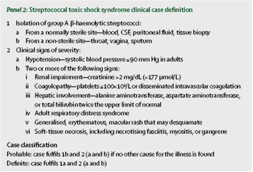
Table 1.
Exotoxins produced by Streptococcus, act as super-antigens causing T-cells activation and cytokine release. M protein, a constituent of the cell wall of streptococcal serotype M1 and M3, is released from the bacterial surface. It is antiphagocytic and has affinity to bind to fibrinogen and forms huge aggregates in blood and tissues. These fibrinogen–M protein aggregates damage endothelium. These mechanisms eventually lead to vascular leakage and hypercoagulability, which in turn cause hypotension, disseminated intravascular coagulation, and multi-organ damage that are characteristic of the streptococcal toxic shock syndrome.
Besides supplementary action to bactericidal effect of penicillin G, clindamycin is also believed to directly inhibit the streptococcal toxin production and facilitate phagocytosis by inhibiting bacterial synthesis of antiphagocytic M protein. The efficacy of clindamycin is unaffected by the bacterial growth phase.
Small case studies and a prematurely terminated RCT found improved survival in patients with streptococcal toxic shock syndrome treated with IVIg. It is thought to provide direct neutralization of streptococcal toxins and to exert immunomodulatory effects on T cells. IVIg can be considered for invasive streptococcal disease when adequate antimicrobials, source control, and sepsis management failed to elicit a response. Guideline suggests an initial dose of 1g/kg on day 1, followed by 0.5g/kg on days 2 and 3.
Hyperbaric oxygen therapy may be beneficial in treating the infection as supported by some case reports.
References
1. E. Lappin, A. Ferguson. Gram-positive toxic shock syndromes. The Lancet Infectious Diseases. 2009 May; 9(5):281-290.
2. Filbin MR, Ring DC, Wessels MR, et al. Case records of the Massachusetts General Hospital. Case 2-2009. A 25-year-old man with pain and swelling of the right hand and hypotension. N Engl J Med 2009;360:281-90.
3. Eric J.B. The molecular basis of streptococcal toxic shock syndrome. N Engl J Med 2004; 350:2093-94.
4. Darenberg J et al. Intravenous immunoglobulin G therapy in streptococcal toxic shock syndrome: A European randomized, double-blinded, placebo-controlled trial. Clin Inf Dis 2003; 37: 333-340.
5. Boyer A., Vargas F., Coste F., et al. Influence of surgical treatment timing on mortality from necrotizing soft tissue infections requiring intensive care management. Intensive Care Med. 2009 May; 35(5):847-53.
6. Mandell, Bennett, & Dolin: Principles and Practice of Infectious Diseases, 6th ed.
7. Schrage B, Duan G, Yang LP, Fraser JD, Proft T. Different preparations of intravenous immunoglobulin vary in their efficacy to neutralize streptococcal superantigens: implications for treatment of streptococcal toxic shock syndrome. Clin Infect Dis2006 Sep 15;43(6):743-6.