
What are the radiological signs present?
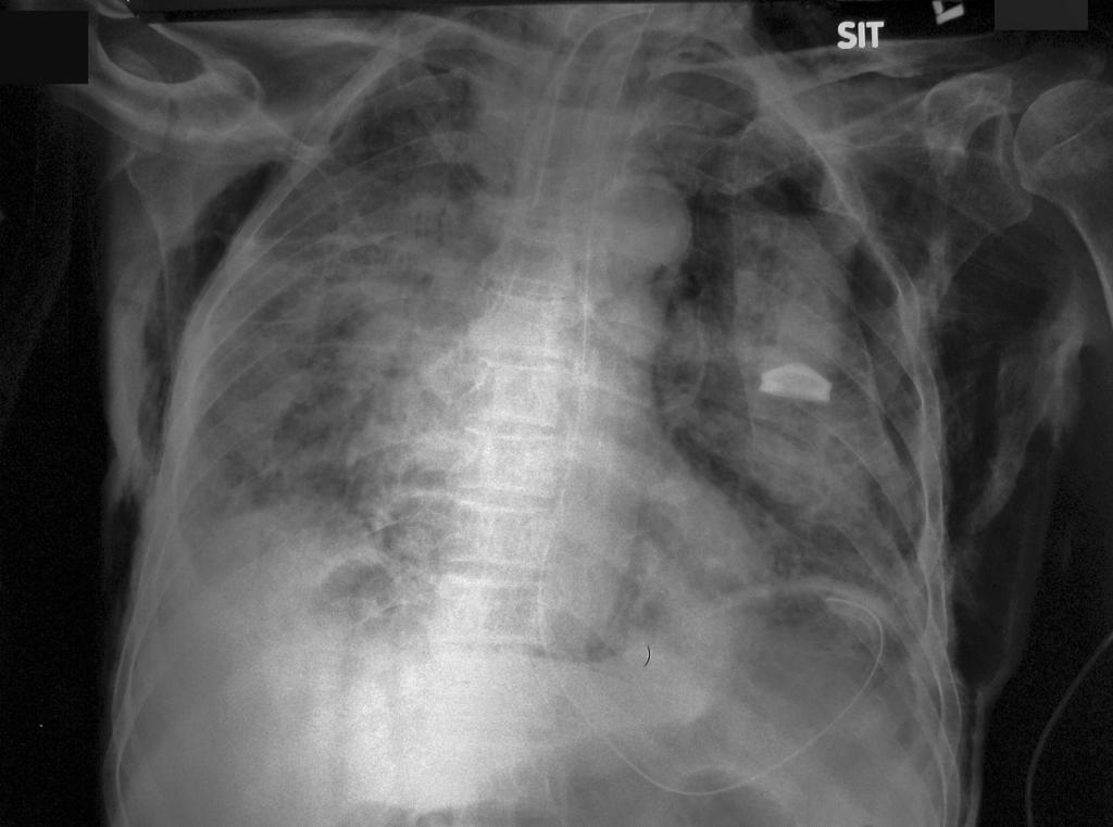
Figure 1
The above chest radiograph shows:
1. bilateral lung infiltrates
2. left pneumothorax
3. extensive subcutaneous emphysema
4. pneumomediastinum with the continuous diaphragm sign
5. Tracheostomy tube and nasogastric tube in-situ
Another chest radiograph of the same patien which shows more clearly the continuous diaphragm sign.
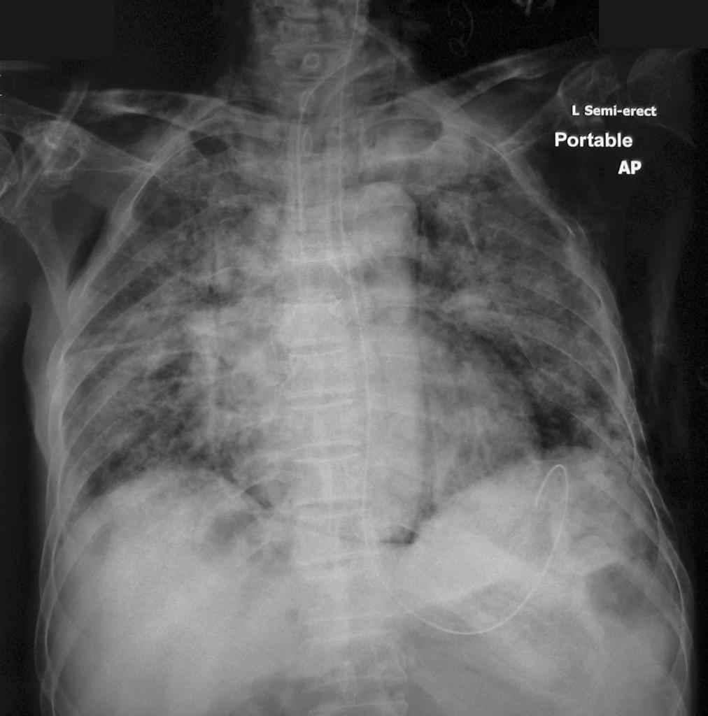
Figure 2
The “continuous diaphragm sign” is produced when a stripe of air is present between the pericardium and the diaphragm. It was first reported in 1973 by Levin. This sign is more readily seen when the patient is examined in the supine position. It is only when there is no overlap of heart on the dome of the diaphragm that the sign will be seen clearly, so the sign is clearer on Figure 2 than on Figure 1.
Reference
1. Levin, B., Clinical Radiology, 1973, 24, 337.
2. P MacPherson and J S DavidsonLetter: Continuous diaphragm sign of pneumomediastinum.Br Med J. 1974 April 13; 2(5910): 119–120. (fulltext available at BMJ here)