
Submitted by Dr CHING Chi Keung, Tseung Kwan O Hospital, Hong Kong, on 2 June 2009
A 52-year-old female had history of hypertension and benign uterine tumour with TAHBSO done in a private hospital in 2004. She was admitted for dyspnoea, cough and exertional chest discomfort for 4 days. There was no ankle edema, fever, chills or rigors. JVP was elevated and there were bilateral basal crackles on auscultation. No murmur was heard. A 2cm soft tissue was noted over lateral aspect of right adductor muscle. SpO2 was 92% room air. Chest x-ray showed pulmonary edema and echocardiography was performed.
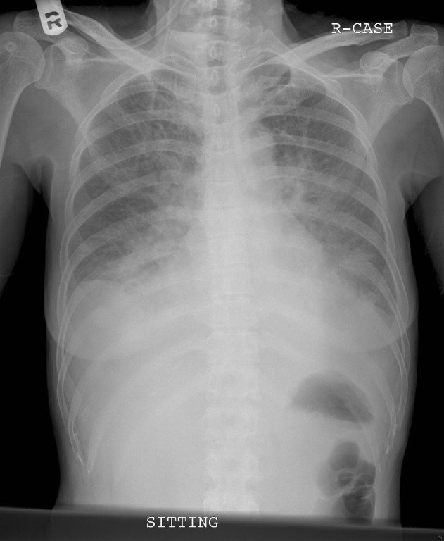
Click Read More to read further.
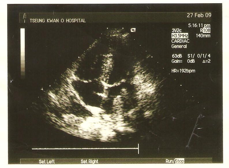
She was transferred to the intensive care unit and CT thorax with contrast was subsequently performed.
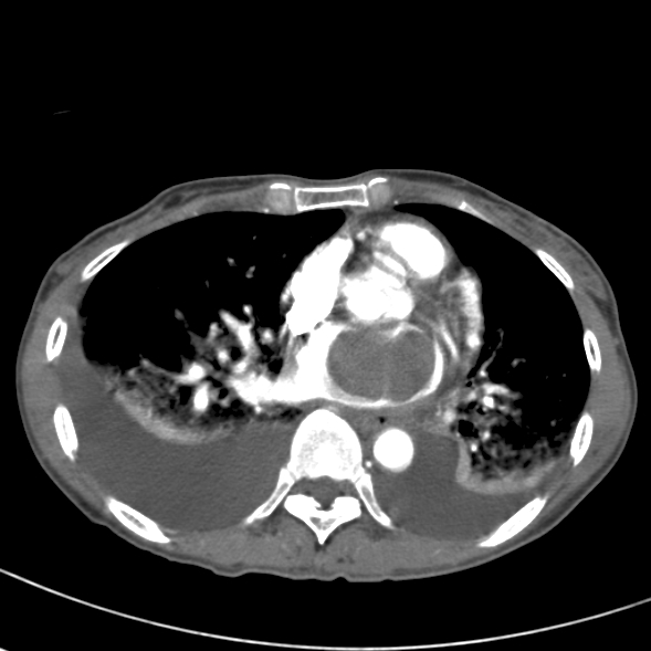
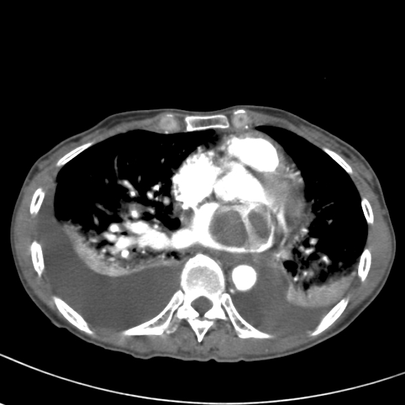
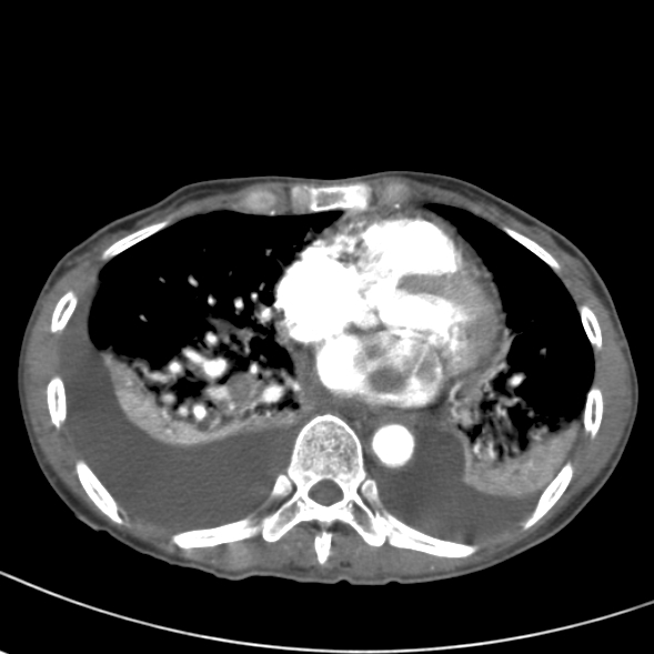
What abnormalities are shown?
Answer
Bedside echocardiograhy showed a left atrial mass attaching to the left atrial wall with severe MR and TR. LVEF was about 60%. CT thorax with contrast revealed a large filling defect at the left atrium measuring 3cm x 4cm x 5cm in size. Another soft density lesion was noted at the region of a branch of the right inferior pulmonary vein. There was also associated bilateral pleural effusion. Impression was left atrial lesion with outflow obstruction resulting in pulmonary edema.
Patient was transferred to the Cardiothoracic Unit of Queen Elizabeth Hospital on the same day. Emergency left atriotomy was performed in view of the risk of decompensation and embolism. During operation, there was a 5 cm firm mass with smooth surface over the LA with base attached to the posterior wall. There were also suspected tumour foci over the orifices of LSPV and RSPV, 1 cm tumour like mass near the annulus with suspected extension to the posterior leaflet and foci near annulus. The anterior leaflet was normal looking. Debulking resection was done and biopsy showed high grade sacroma consistent with leiomyosarcoma.
Slide review of the surgical specimen of TAHBSO in 2004 showed leiomyoma. Repeat CT thorax, abdomen and pelvis about 2 months later showed enhancing soft tissue mass in the left atrium extending to the left pulmonary vein which was compatible with the history of leiomyosarcoma. A 0.6 cm nodule suspicious of malignancy was noted at the lower lobe of left lung. Two hypodense lesions measuring about 1.4 cm were noted at segment 7 of the liver. Proper liver CT or MRI may be justified. She was then referred to the Department of Clinical Oncology for chemotherapy.
The right thigh mass was assessed by orthopaedic surgeon and MRI of both upper thighs showed only small apparent cystic lesion along the femoral origin of the right adductor muscle. Follow-up USG to confirm the cystic nature of lesion may be helpful.
Virtually all types of sarcoma have been reported in the heart. Diagnostic approach replies upon echocardiography, CT or MRI to define the presence of tumour and its anatomic relationship to normal structures. The most frequently described sarcomas include angiosarcomas, rhadomyosarcomas, fibrosarcomas and leimyosarcoma. They have a high rate of local recurrence and systemic spread. Most patients develop recurrent disease or die of malignancy even if the tumour can be completely resected. Adjuvant chemotherapy has been used to improve the poor result with resection alone.