
Submitted by Dr LAU Chun Wing Arthur on 10 April 2009
ICU, Pamela Youde Nethersole Eastern Hospital, Hong Kong
A M/45 was admitted for post-op care in the ICU for an elective right nephrectomy. What was the indication?
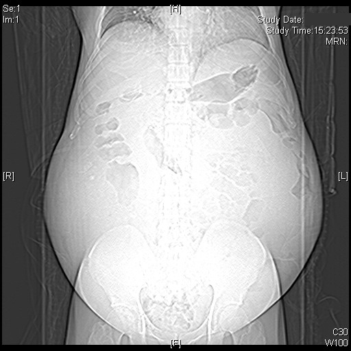
Figure. CT scan done a year ago.
Click read more to see the answer.
See more images:
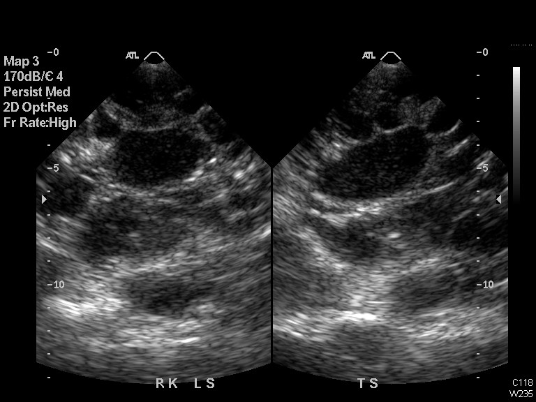
Fig. USG right kidneys (longitudinal and transverse sections) done a year ago.
| {morfeo 28} |
CT abdomen and pelvis images
Answer
This patient had autosomal dominant polycystic kidney disease (ADPKD) with end-stage renal failure on hemodialysis. Because of the huge huge polycystic kidneys, right nephrectomy had to be done to create room before consideration of potential kidney transplantation.
US ABDOMEN: 1. Polycystic kidney disease. 1.4cm x 1.9cm x 2cm echogenic nodule in the upper pole of left kidney. Complex cystic lesions in both kidneys, which may represent previous hemorrhage or inflammation. In view of clinical history of gross haematuria, the possibility of malignancy cannot be excluded. Suggest further evaluation as clinically indicated. 2. Multiple liver cysts. Tiny 6.5mm echogenic nodule in the right lobe of liver; nature is uncertain.
CT SCAN OF ABDOMEN AND PELVIS: Enlarged both kidneys with multiple cysts, many with calcified wall and some of these are slightly hyperdense, probably related to high proteinaceous content or previous hemorrhage. Appearance is consistent with polycystic renal disease. Multiple liver cysts. Mild ascites in pelvic cavity.
Discussion
A retrospective Hong Kong autopsy study published in 1993 of 41 Chinese patients who had an autopsy diagnosis of APKD showed that nineteen (46%) patients reached end-stage renal disease (ESRD) at the time of death. Ruptured berry aneurysm caused death in 3 patients while 3 others had spontaneous intracerebral hemorrhage. Left ventricular hypertrophy was found in 24 (59%) patients. Polycystic liver was present in 26 (63%) patients. This frequency was found to be higher than the 4% to 60% (average 47%) reported in similar studies conducted in Western countries. The probability of patients being alive and without ESRD, estimated using a life table analysis, was 83%, 54%, 32% and 12% by the age of 40, 50, 60 and 70 respectively.
My colleague in the Department of Medicine of Pamela Youde Nethersole Hospital, Dr LO Hok King Stanley, now Specialist in Nephrology, also provided some local data in his 2001 Nephrology Exit Dissertation (free and openly availabe at the Hong Kong College of Physican website here) The main points are summarized as below:
Autosomal dominant polycystic kidney disease (ADPKD) patients followed up in a regional hospital (PYNEH) were reviewed and ADPKD was estimated to have a prevalence of 99 per 1,000,000 populations with a mean age of 51.8 years in the Eastern Hong Kong Island, accounting for 1.4% of the corresponding dialysis populations.
The following factors were more likely related to normal function:
1. patients younger than 50 years (p=0.016; Odd ratio=3.62; 95% C.I.= 1.24-10.6)
2. having a positive family history of ADPKD (p=0.001; odd ratio= 8.25; 95% C.I.=2.27-30.0)
3. proteinuria less than 0.5g/day (p=0.028; odd ratio=9; 95% C.I.=1.002-80.8)
4. kidney size less than 15 cm (p=0.055)
Patients with normal renal function also had low risk of left ventricular hypertrophy (p=0.03; odd ratio=0.12; 95% C.I.=0.014-1.06).
Slower renal function decline was more likely to be found in:
1. patients younger than 50 years (p=0.03, odd ratio=3.51; 95%C.I.=1.09-11.3)
2. in those with a family history of ADPKD (p=0.031; odd ratio=3.71; 95%C.I.=1.09-12.6)
3. in those with normal renal function (p=0.021, odd ratio=4.4; 95% C.I.=1.22-15.8)
Controls of blood pressure in 64.4% patients at outpatient clinic were not satisfactory according to the recommendation of Sixth Joint National Committee. Blood pressure control should be improved.
Review of the contemporary literature on ADPKD showed that the following factors were poor prognostic factor for renal progression:
1. younger age at diagnosis
2. male gender
3. black race
4. PKD-1 and ACE DD genotype
5. history of gross haematuria
6. urinary tract infection in male
7. proteinuria
8. bigger kidney size
9. poor blood pressure control
Angiotensin converting enzyme inhibitor was shown to be advantageous over other anti-hypertensives. Renal diet is of limited benefit for renal progression. Family screening is important to identify at-risk patients for hypertension and monitoring of renal function. Those with family members with aneurysm rupture required screening with magnetic resonance angiogram.
There are two types (Types I and II) of autosomal dominant polycystic kidney disease and they have similar pathological and physiological features. Tremendous cystic enlargement of both kidneys like those in the present patient is characteristic of ADPKD. For Type I disease, the aberrant gene is PKD1, with chromosomal abnormality at 16p13.3, which encodes the protein Polycystin-1. Polycystin-1 is a membrane mechanoreceptor-like protein, which interacts with the extracellular environment to regulate renal morphogenesis and differentiation, including control of renal tubule diameter through effects on epithelial-cell proliferation, adhesion, and migration. Type II disease is also autosomal dominant. It has a later onset of symptoms and a slower rate of progression to renal failure, and the abnormality is at the PKD2 gene, the abnormal protein product is polycystin-2. Patients can even have mutations in both the PKD1 and PKD2 genes (transheterozygotes) and they will have a more severe clinical course than those with mutations in only one of the genes. Autosomal recessive polycystic kidney disease is much rarer than the autosomal dominant form, with an incidence of 1 in 20,000 live births, and often causes fetal or neonatal death. Our patient is more compatible with Type I ADPKD.
Pattern of cyst formation is the same in the two types of ADPKD. Cystic outpushings arise in every tubule segment and rapidly close off from the nephron of origin. In autosomal recessive polycystic kidney, the cysts are derived from collecting tubules, but always remain connected to the nephron of origin.
The polycystins regulate tubular and vascular development in the kidneys and other organs (liver, brain, heart, and pancreas), therefore these patients also have liver and pancreatic cysts, and also an increased risk of aortic aneurysms and heart-valve defects, and some kindreds have five times the risk in the general population of sudden death from ruptured intracerebral aneurysms. There was no pancreatic cyst in our patient.
USG criteria have been established for the diagnosis, include: at least two unilateral or bilateral cysts in persons younger than 30 years of age, at least two cysts in each kidney in persons 30 to 59 years of age, and at least four cysts in each kidney in persons 60 years of age or older.
There was an interesting study in 2006 on the volume progression in this condition (CRISP study). It was found that with time the total kidney volume and total cyst volume increased exponentially. The baseline total kidney volume predicted the subsequent rate of increase in volume, independently of age. A baseline total kidney volume above 1500 ml (in 51 patients) was associated with a declining glomerular filtration rate by 4.33±8.07 ml per minute per year. Total kidney volume increased more (in 135 patients) with PKD1 mutations by 245±268 ml than with PKD2 mutations (in 28 patients) by 136±100 ml (P = 0.03).
References
1. Chan KW. Adult polycystic kidney disease in Hong Kong Chinese: an autopsy study. Pathology. 1993 Jul;25(3):229-32.
2. Marcos Lucon, Luiz Estevam Ianhez, Antonio Marmo Lucon, Jose L. Chambô, Emil Sabbaga, Miguel Srougi. BILATERAL NEPHRECTOMY OF HUGE POLYCYSTIC KIDNEYS ASSOCIATED WITH A RECTUS ABDOMINIS DIASTASIS AND UMBILICAL HERNIA. CLINICS 2006;61(6):529-34
– Report of a case with similarly huge polycystic kidneys. Techniques of nephrectomy and umbilical hernia and rectus abdominis diastasis repair were described.
3. Wilson PD. Mechanisms of disease: Polycystic Kidney Disease. N Engl J Med 2004;350:151-64.
Jared J. Grantham, M.D. Autosomal Dominant Polycystic Kidney Disease. N Engl J Med 2008;359:1477-85.
4. Ravine D, Gibson RN, Walker RG, Sheffield LJ, Kincaid-Smith P, Danks DM. Evaluation of ultrasonographic diagnostic
criteria for autosomal dominant polycystic kidney disease 1. Lancet 1994;343:824-7.
5. Grantham JJ, Vicente E. Torres, Arlene B. Chapman, et al, for the CRISP Investigators*. Volume Progression in Polycystic Kidney Disease. N Engl J Med 2006;354:2122-30.
6. Grantham JJ. Autosomal Dominant Polycystic Kidney Disease. N Engl J Med 2008;359:1477-85.
7. LO Hok-King Stanley. POLYCYSTIC KIDNEY DISEASE: AN EXPERIENCE IN A REGIONAL HOSPITAL. Department of Medicine, Pamela Youde Nethersole Eastern Hospital, December 2001 Nephrology Exit Assessment Exercise. Accessed on the Hong Kong College of Physicans website on 11 April 2009.