
Submitted by Dr LAU Chun Wing Arthur on 11 March 2009
Department of Intensive Care, PYNEH
A 59-year-old man of good past health was transferred into the ICU for respiratory failure and shock.
 |
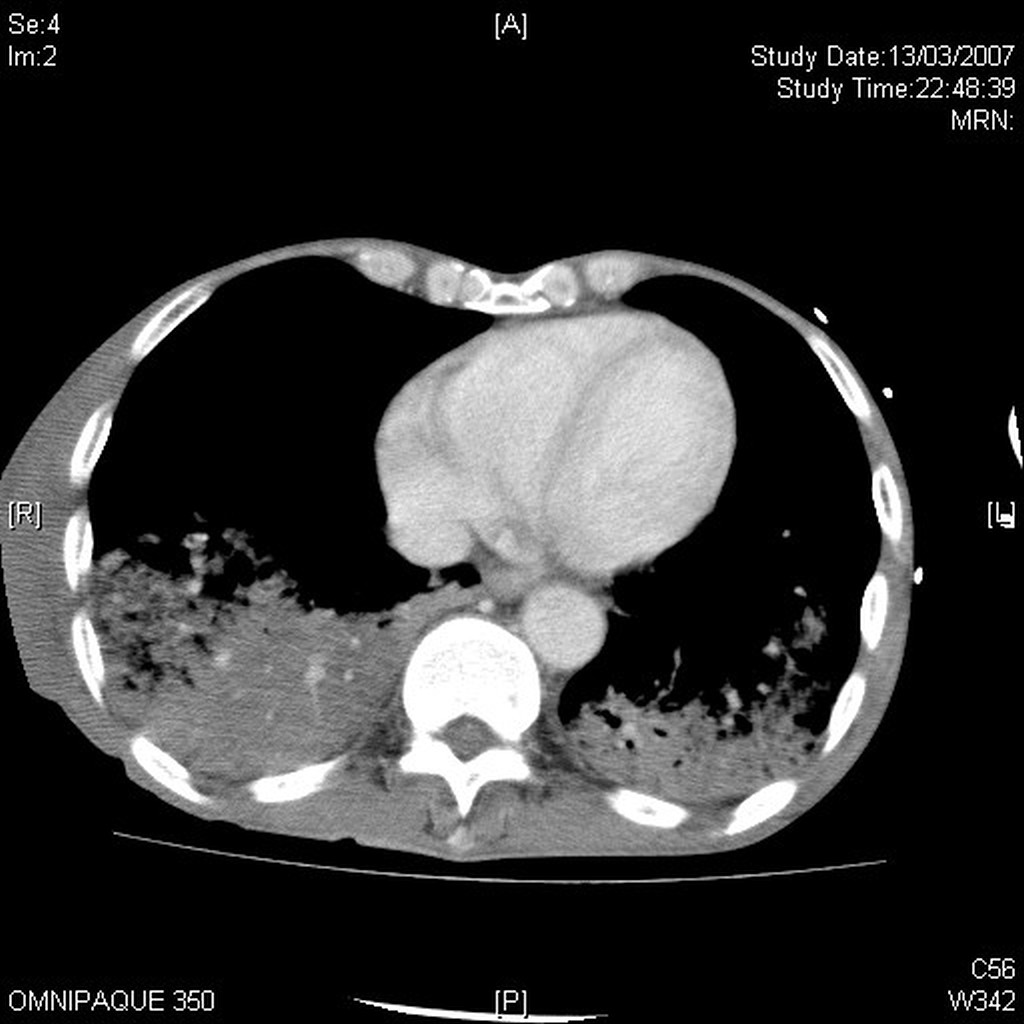
Upper photo: figure 1; Lower figure: figure 2
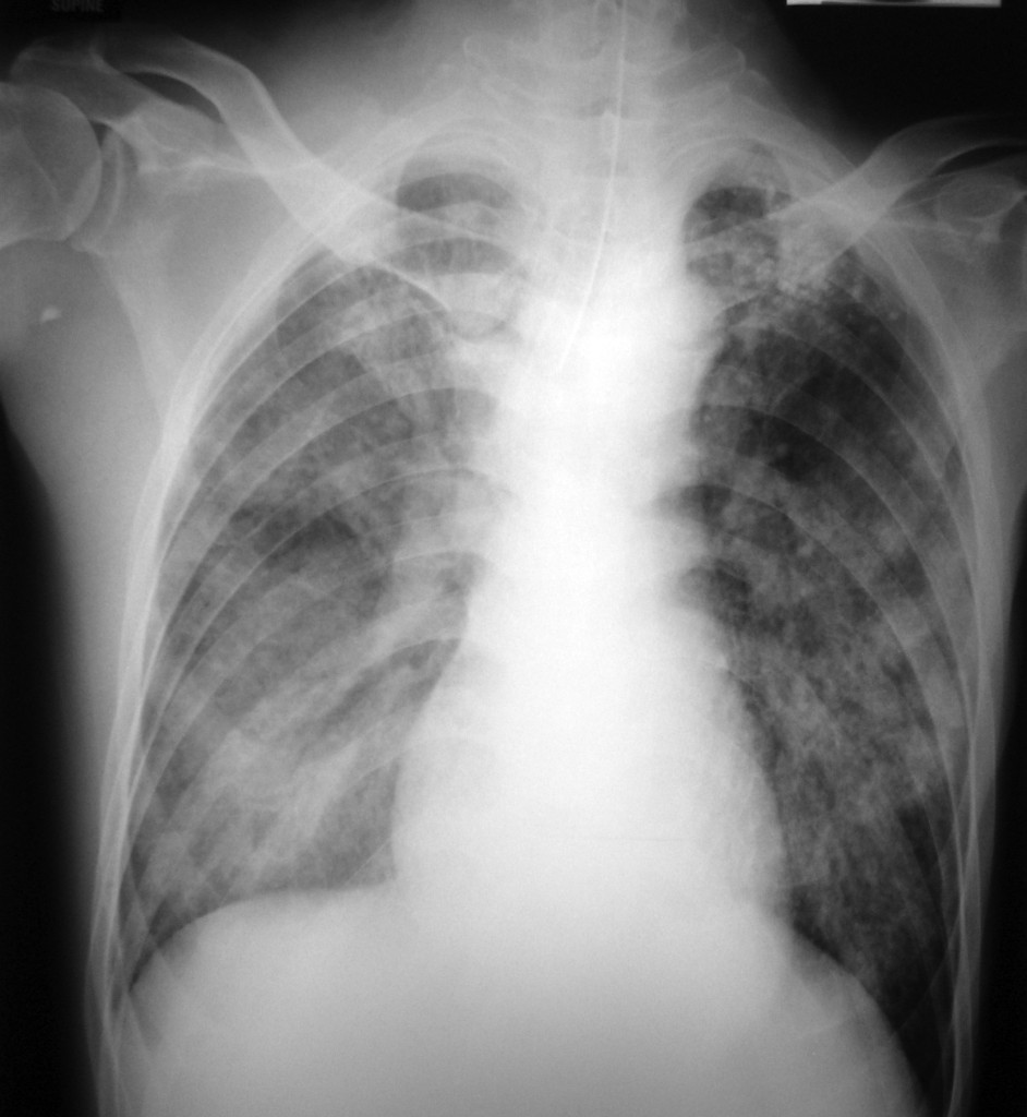
He complained of weight loss and generalised weakness for 1 month. The CXR is shown in Figure 1. Because he developed significant desaturation, he was intubated and mechanically ventilated. He developed severe shock despite fluid resuscitation and high dose vasopressors.
Labs: Derranged LFT: ALP 140, ALT 360, total bilirubin 72; Na 140, Ur 15, Cr 164; WBC 4, Hb 7.4 (retic 1.4%), Plt 59 and deranged clotting c/w DIC
Bedside USG abd – no SOL, biliary tree not dilated
Because of shock and deranged LFT, CT abdomen (Figure 2) was done to see if there was any source of sepsis that could be missed by USG. Report: normal excpet small amount of ascites, bilateral lower lobe consolidations are noted, more on right side.
Sputum for AFB smear was negative. Could it be passed as just a case of severe bacterial pneumonia? How would you confirm the diagnosis?
On reviewing the images, it was found that the CXR showed not just simple diffuse consolidative changes. Multiple lung nodules extending to the lung peripheries could be seen on close-up (Figure 3), but which was not easily picked up by the initial over-penetrated CXR (not shown). There were also fibrocalcifications over the left upper lobe suggestive of old TB changes.
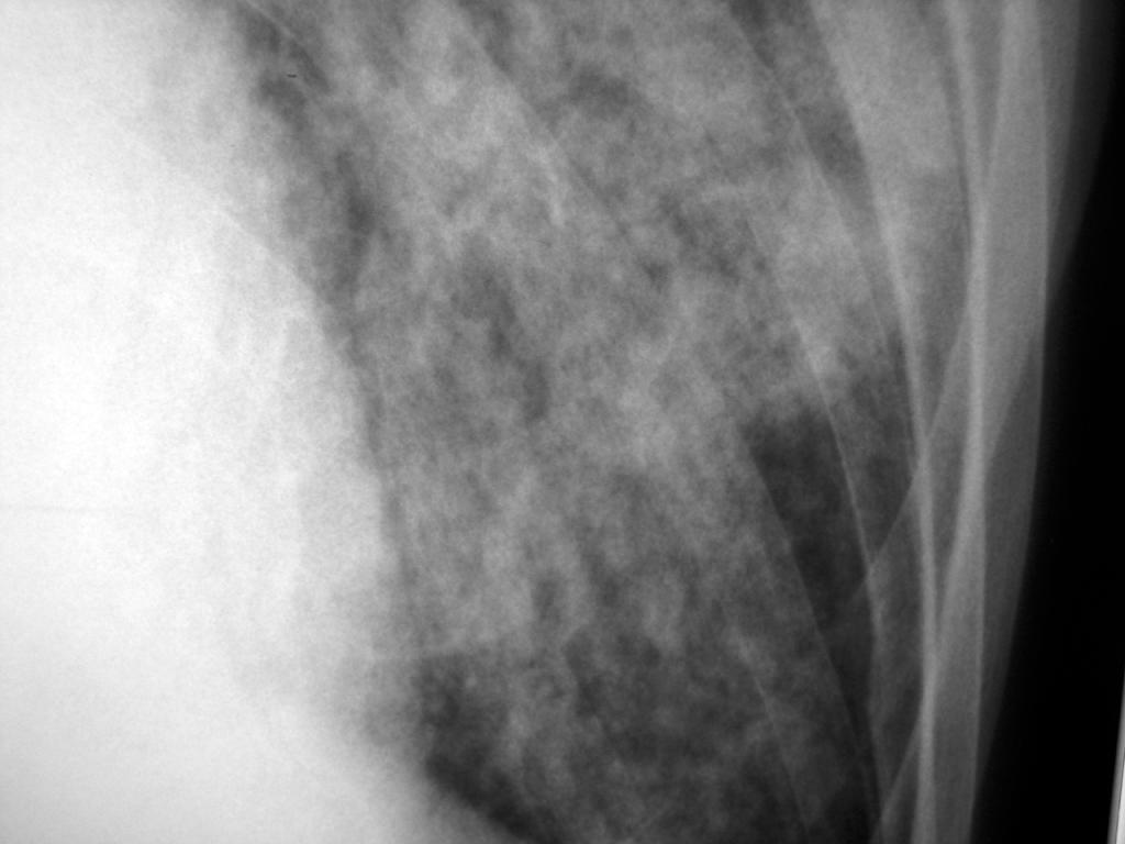
Figure 3
Because of the above findings, the CT abdomen was reviewed. Using the radiological imaging function on the Centricity software on the Electronic Patient Record (ePR) system, we changed the CT abdomen to lung parenchyma window, and looked at the bases of both lungs.
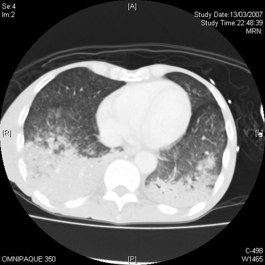
Figure 4. A CT meant for abdominal exam changed to lung parenchymal windows to inspect the lung bases.
As the CT is not 1 mm thin cuts as done for high-resolution CT thorax, a very clear texture of the lung parenchymal could not be appreciated. However, with the images obtained by CT machines nowadays, thin images can always be retrieved and reconstructed retrospectively by consultation with the radiologist. Both diffuse tiny lung nodules and consolidative changes could be seen in the lung parenchyma.
Taking into account the almost pancytopaenic picture, we believed disseminated TB could be the diagnosis, so a bone marrow aspirate and trephine biopsy (Figure 5) were done. Acid-fast bacilli were found in the bone marrow.
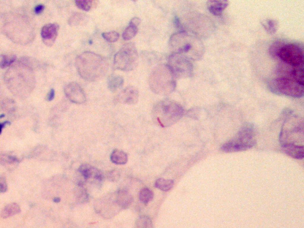
Figure 5
Bone marrow aspirate for AFB was negative, but rapid AFB culture was positive, and DNA probe showed the presence of M. tuberculosis complex rRNA. Marrow trephine sections showed small amount of marrow tissue with presence of trilineage haematopoiesis, with some hypocellular areas showing gelatinous transformation. No abnormal infiltrations were seen. Several histiocytic aggregates were noted. ZN stain showed several AFB. There was no increase in the amount of reticulin fibres. Sputum for AFB smear was negative, but was AFB culture positive for M. tuberculsosis. The mycobacteria was sensitive to streptomycin, rifampicin and ethambutol, but was resistant to isoniazid (pattern same in sputum as in the bone marrow). The rate of monoresistance to isoniazid in new case (i.e. not previously treated) was 2.09% (Figure 6) locally in Hong Kong.
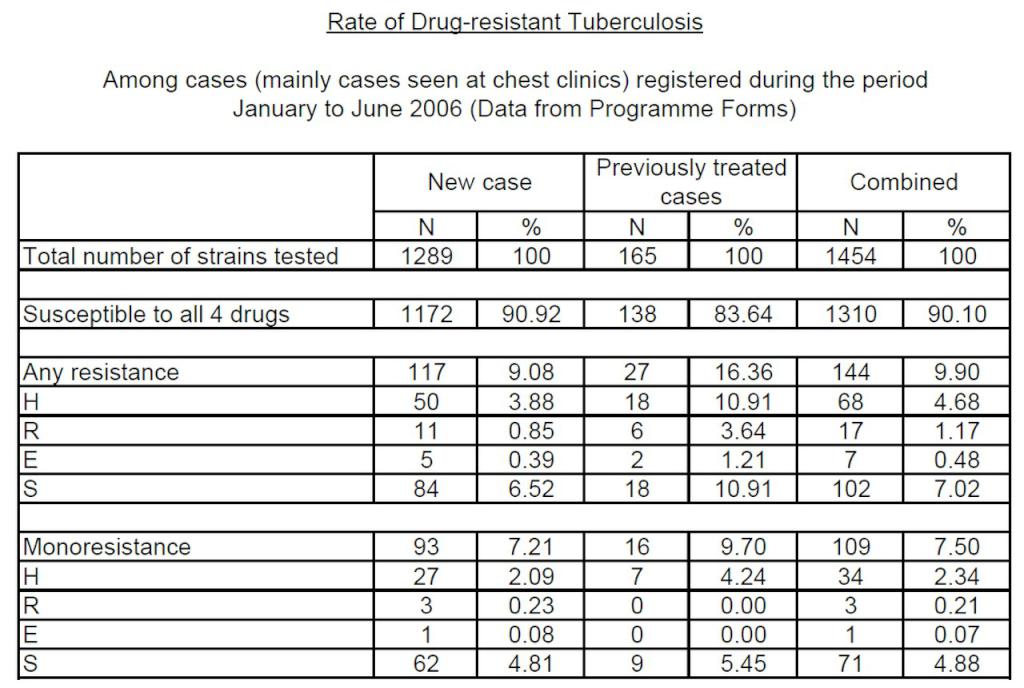
Figure 6. Rate of drug-resistant TB in Hong Kong (ref: Annual Report 2006, TB and Chest Service of the Department of Health, Hong Kong)
The patient had actually been started empirically on anti-tuberculosis medications even before the diagnosis of disseminated TB was made. However, because of the significant liver and renal functions derangement, only levofloxacin and ethambutol could possibly be prescribed. He eventually succumbed despite maximal support.
Learning points
1. Read and interpret CXR carefully. Know the difference between alveolar (consolidative) and interstitial (honeycomb or nodular or miliary) changes and correlate with the overall clinical picture. This patient actually had both. Mind the limitations of an over-penetrated film. The Picture Archiving and Communication System (PACS), i.e. filmless radiology system, may help reduce this drawback if we care to adjust brightness and contrast.
2. Miliary tuberculosis (or disseminated TB) is a form of tuberculosis that is characterized by a wide dissemination of TB into the human body. Its name comes from a distinctive pattern seen on a CXR of many tiny spots (1 – 5 mm) distributed throughout the lung fields like millet seeds, thus the term “miliary” tuberculosis. It is caused by (a) erosion of the infection into a pulmonary vein, and then the bacteria reach the left side of the heart and enter the systemic circulation and seed many other organs such as the liver and spleen, and/or (b) the bacteria may enter the lymph node, drain into a systemic vein and eventually reach the right side of the heart, from the right side of the heart, the bacteria may then seed the lungs, causing the “miliary” appearance, or futher re-seed other organs as in a.
3. Overall sputum for AFB positivity rate is only around 50% in TB patients. Rate may be higher for cavitary changes or extensive parenchymal changes like TB pneumonia (usually sputum AFB positive, i.e. open TB, therefore more infectious), but will be negative in miliary TB (sputum AFB negative, i.e. not open, or not infectious to people around).
4. It is very common, but not correct, to label all nodules seen on a TB CXR as miliary, but in fact, only those 1 to 5 mm in size can be called miliary. This has clinical implication, as explained above. For this patient, the extremely small nodules on the CT should represent miliary nodules, while the larger and confluent ones should represent cosolidative changes.
5. There are much more to see on a CT than the report alone. Change the window to discover more, or ask your radiologist for help if necessary.
6. You can protect yourself and your staff around if you can correctly interpret the CXR. TB is air-borne!