
Submitted by Dr LAU Chun Wing Arthur on 26 Feb 2009
Department of Intensive Care, PYNEH
This 51-year-old lady had influenza A pneumonitis complicated with ventilator-associated pneumonia. She developed persistent atrial fibrillation, abdominal distension and worsening septic shock. CT abdomen and pelvis was done.
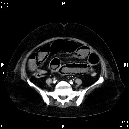
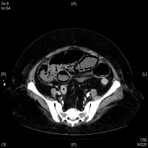
What are the ddx of this condition? Click here to read more.
CT abdomen and pelvis with contrast findings: Numerous dependent tiny bubbly lucencies are seen over the dependent portion of the jejunum and ileum, which are suspicious of pneumatosis intestinalis. The small bowel is distended. Bowel wall enhancement is still noted in the proximal jejunum but no definite wall enhancement is seen in part of the small bowel, probably ileum. No definite portal venous gas is seen. The celiac trunk, main SMA & SMV appear patent with no definite filling defect but the opacification of the branches of the SMA is not well appreciated due to their small size. Overall features can be due to bowel ischaemia, infection or other pathologies. No significant colonic distension is noted. Two hypoenhancing lesions are noted in both lobes of the liver, up to about 3mm in size, probably cysts. Otherwise, no focal hepatic mass lesion is noted. Portal and hepatic veins are normally enhanced. Intrahepatic ducts and common bile duct are not dilated. A 4mm hyperdense gallbladder stone is seen. The spleen, pancreas and both adrenals are unremarkable. Both kidneys appear unremarkable and no hydronephrosis is noted. A large lobulated heterogenous enhancing mass measuring about 7.0cm X 3.4cm x 3.2cm is noted over the uterus, probably a uterine fibroid. No gross ascites is noted. Impression: suspicious of pneumatosis coli. The small bowel is distended. Bowel wall enhancement is still noted in the proximal jejunum but no definite wall enhancement is noted in part of the small bowel, probably ileum. The celiac trunk, main SMA & SMV appear patent with no definite filling defect. Overall features can be due to bowel ischaemia, infection or other pathologies.
Laparotomy, dx – Acute ischaemic bowel: Extensive small bowel infarct from proximal jejunum to ileocaecal junction, multiple large perforations due to bowel wall necrosis over ileum (wrapped around by omentum over right side of abdomen), large mesenteric vessels over mesentery – pulse palpable, small vessels over mesenteric arcade and distal to arcade – no pulse palpable
Discussion
Pneumatosis intestinalis is defined as the presence of gas in the wall of the gastrointestinal tract, which can affect any part except the esophagus. Two main theories have been proposed: 1. A mechanical theory hypothesizes that gas dissects into the bowel wall from either the intestinal lumen or the lungs via the mediastinum due to some mechanism causing increased pressure (e.g. in asthma, emphysema, mechanical ventilation); 2. A bacterial theory proposes that gas-forming bacilli enter the submucosa through mucosal rents or increased mucosal permeability and produce gas within the bowel wall (as in our patient). Studies have shown that gas collections in the bowel wall can have a hydrogen content of up to 50%. Hydrogen is a product of bacterial metabolism and is not produced by human cells. The presence of gas in the mesenteric or portal venous system in mesenteric ischemia is indicative of an unfavorable prognosis.
Further information on the web:
1. Ho LM et al, AJR 2007: lots of radiological images and a table of differential diagnoses
2. Brigham RAD: gross pathology specimen