
Submitted by Pablo Blanco, José Luis do Pico on 4 Jan 2012
Pablo Blanco, Critical care physician, Intensive Care Unit, Dr. Emilio Ferreyra Hospital, 59 Avenue 4801. Necochea, Argentina. Email: ohtusabes@gmail.com
José Luis do Pico, Critical care physician, FCCM, Intensive Care Unit, Dr. Emilio Ferreyra Hospital, 59 Avenue 4801. Necochea, Argentina. Email: dopiconec@gmail.com
Correspondence to: dopiconec@gmail.com
ABSTRACT
Introduction: Although this is not the first case published of symmetrical peripheral gangrene, we hypothesize that the rational management of the clinical conditions associated with symmetrical peripheral gangrene must prevent, reverse or attenuate the clinical picture and thus reduces morbidity and mortality.
Case presentation: 34 years old male patient with a personal history of alcoholism presented with severe sepsis from a right lung abscess. He developed disseminated intravascular coagulation, requires high doses of noradrenaline infusion and presented acral gangrene in the four extremities. Finally died.
Conclusion and Review: The clinical case resolution shows various tips not only about symmetrical peripheral gangrene but yes about other very important management points accordingly with patient suffering, ie: Acute respiratory distress syndrome and prone position, alveolar recruitment maneuvers, disseminated intravascular coagulation criteria, etc. The management of symmetrical peripheral gangrene is multidisciplinary, for that, the clinical impact of the present clinical case is broader across medicine.
Keywords: severe sepsis, lung abscess, symmetrical peripheral gangrene, noradrenaline infusion, disseminated intravascular coagulation.
A 34-year-old male patient was admitted to the emergency department suffering from respiratory distress. He was lucid, cooperative and was showing a significant increase in his respiratory effort. He had been experiencing progressive dyspnea at rest for the previous seven days as well as mucopurulent expectoration. He had condensation semiology throughout the right hemithorax and at the base of the left hemithorax. Poor peripheral perfusion. He was diaphoretic and feverless.
Front chest x-ray: non-homogeneous infiltrate with air bronchogram at the base of the left lung. Thick wall cavitated image with air-fluid level spread throughout practically the whole right lung field. (Figure 1, see Appendix).
Thoracic CT Scan without intravenous contrast: image with air-fluid level spread throughout practically the whole right lung field. Non-homogeneous infiltrate with air bronchogram in posterior segments of the lower left lobe. (Figure 2, see Appendix).
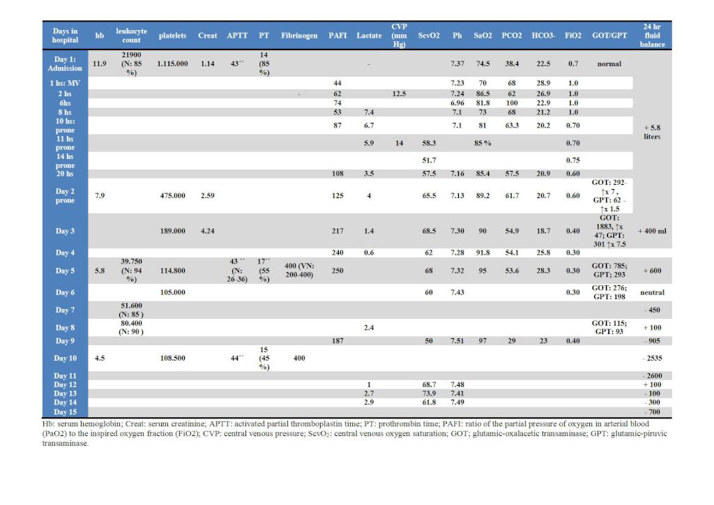
Blood analytical review at admission: Hemoglobin: 11.9 gr/l, leukocyte count per mm3: 21.900 (neutrophils: 85%), potassemia: 2.3 meq/l; platelet count per mm3: 1.110.000¸ prothrombin time (PT): 14 seconds (86%); activated partial thromboplastin time (APTT): 43 seconds; arterial pH: 7.37; partial pressure of oxygen in arterial blood (PaO2): 40 mmHg, fraction of inspired oxygen (FiO2): 70%; partial pressure of carbon dioxide in arterial blood (PaCO2): 38.4 mmHg, bicarbonate (HCO3-): 22.5 mmol/l, oxygen saturation in arterial blood (SaO2): 74.5 %, PaO2/FiO2 (PAFI): 57. (Table 1, See Appendix).
Electrocardiogram: sinus rhythm, 120 beats per minute, PR interval: 0.14 seconds, diffuse flattening of T waves, corrected QT interval: 0.44 seconds.
During admission at the Intensive Care Unit (ICU) orotracheal intubation and protective mechanical ventilation (MV) was started. Persistent, refractory hypoxemia in spite of the maneuvers used to increase mean airway pressure (See Appendix: alveolar recruitment maneuver used). Refractory arterial hypotension to isotonic crystalloid fluids, 14 mmHg of central venous pressure (CVP) were reached. Therefore, treatment began with an 0.8 microgram per kilogram per minute (ug/kg/min) noradrenaline dose to achieve mean arterial pressure (MAP) equal to or higher than 65 mmHg. Intravenous potassium replacement was started. A right anterolateral thoracotomy was performed by surgical team as a result of a suspected purulent pleuro-pulmonary collection. An empty pleural cavity was confirmed as well as lung bulging at middle lobe level, where 2 liters of purulent material was obtained. Two drainages were placed. He returns from the operating room experiencing cardiac arrest in pulseless electrical activity. Cardiopulmonary resuscitation was performed and the patient recovered pulses 6 minutes later. A total dose of 2 mg of adrenaline was administered. Two blood cultures were taken before antimicrobial starting (ampicillin-sulbactam and clarithromicyn). A 20 gauge catheter was placed for invasive measurement of blood pressure in left radial artery. There was persistent refractory hypoxemia and therefore was placed in a prone decubitus position. Considering arterial lactate and central venous oxygen saturation (ScvO2) values a 4 ug/kg/min dose of dobutamine infusion was started. The patient required noradrenaline increases up to 3 ug/kg/min for several hours to maintain MAP goal. Intravenous hydrocortisone was added at stress doses. Anuric.
Thoracic drainages oscillation with bubbling in both times.
20 hours after being hospitalized the platelet count was 475.000 per mm3, serum creatinine increased 2.3 times and hepatic enzymes also increased: serum Glutamic-oxalacetic transaminase (GOT) times 7 and serum Glutamic-piruvic transaminase (GPT) times 1.5, both compared to the values at the moment of admission. (table 1, see Appendix).
Second day in hospital: Good central perfusion with cold hands and feet. Slow and insidious onset of cyanosis, gradually increasing. Decreased amplitude to palpation of the bilateral radial, dorsal pedis and posterior tibialis pulses. Dobutamine was interrupted. A 2000 ml isotonic crystalloid fluid challenge was performed. CVP 14 mmHg. The invasive blood pressure line stopped working suddenly. The catheter was not bent nor occluded with bubbles, blood or clots. Right femoral invasive blood pressure line was placed. The noradrenaline dose was reduced to 1.8 ug/kg/min. Dobutamine was resumed as a result of lactate increase and ScvO2 reduction. The patient was kept in a prone decubitus position for approximately 14 hours.
Third day in hospital: significant improvement in oxygenation gasometric parameters that was maintained in dorsal decubitus position.
Noradrenaline dose: 0.9 ug/kg/min. Dobutamine stopped.
Serum creatinine value increased 3.7 times.
Beginning of renal support therapies (RST): intermittent daily hemodialysis (IDHD) by means of left femoral double-lumen catheter. The patient was not anticoagulated during the dialytic procedure. Good hemodynamic tolerance during two hours hemodialysis. Not ultrafiltrated volume (UF).
Lung collection culture: Group F Streptococcus. Sensitive to: Clindamycin, Cefepime, Erithromicine, Ceftriaxone. Clindamycin was added to the antibiotic treatment.
Negative blood cultures.
Negative Hepatitis B and Human Immunodeficiency Virus blood tests.
Poor evolution of lesions in the distal third of the four extremities with a significant increase in coldness and cyanosis, limited to hand and feet roots (“glove” and sock” as appropriate). No radial, dorsal pedis and bilateral posterior tibialis pulses are felt.
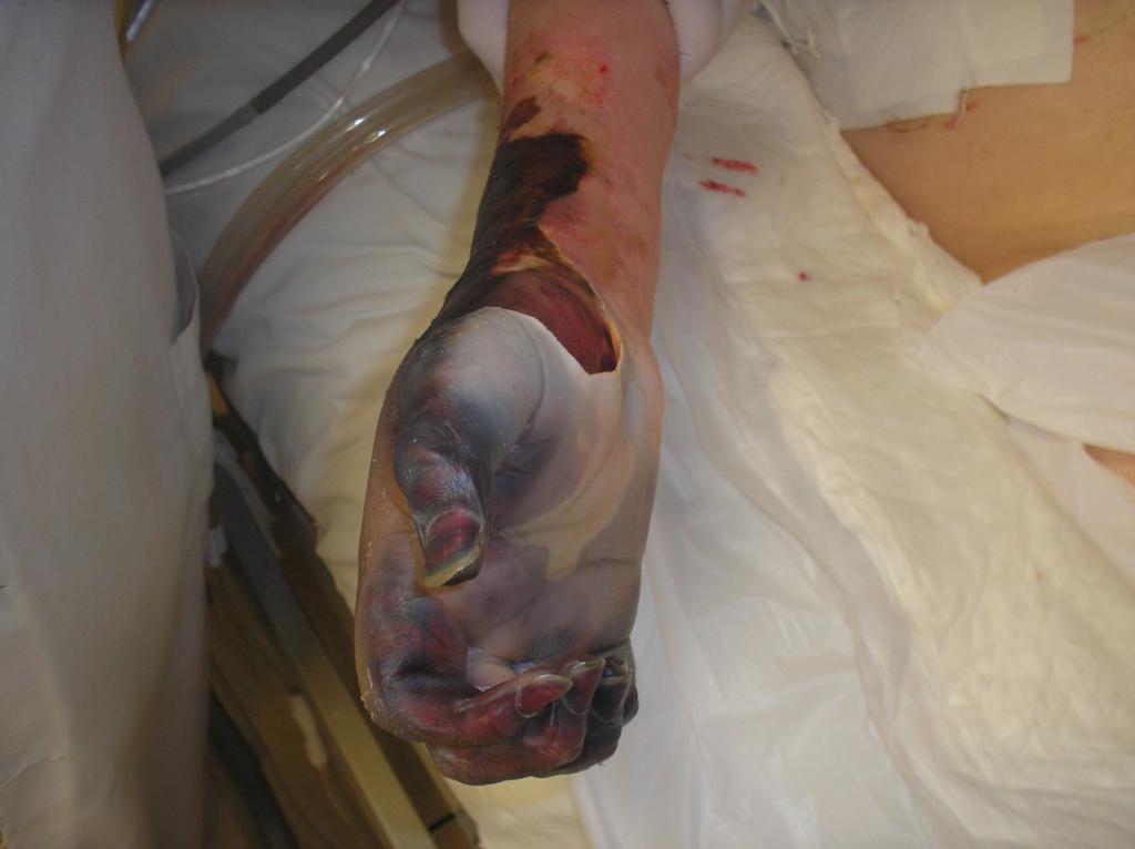
Fourth day in hospital: fewer amounts of noradrenaline required (0.6 ug/kg/min). Nutritional support (NS) began with polymeric diet. Two hours hemodyalisis without complications. UF: 700 ml. Sedoanalgesia was interrupted. The coldness and cyanosis of the distal areas of the extremities became dark brown and hardened lesions. The skin was stiff and phlyctenas with serohaematic, necrotic material. Perfectly restricted to “glove and sock”. (figures 3,4 and 5, Appendix).
Fifth day in hospital: No vasoactive medication needed. The patient did not awake yet. Still anuric. IDHD without complications. PAFI 212.
The lesions in the extremities became dark black and drier, compatible with the onset of dry gangrene (figures 6 to 12, Appendix).
Seventh day in hospital: 50% drop in hepatic enzymes (Appendix). No spontaneous eye opening. Showed signs of pain when abdominal palpation was performed. The ultrasonographic abdominal study showed absence of free fluid in the cavity and distended intestinal loops with fluid content, without peristaltic movements. Intestinal ischemia was suspected and NS was interrupted. Still anuric. Poor hemodynamic tolerance to two hours hemodyalisis. UF: 1400 ml.
Ninth day in hospital: generalized flaccidity. Eye opening as result of pain. The lesions in the extremities had a drier aspect. The thrombosed venous network was visible and his phalanges were stiff and had changed into a lighter color with a translucent aspect (See Appendix). Two hour hemodyalisis, UF: 1500 ml, well tolerated.
Tenth day in hospital: tense abdomen, gestures of pain when palpated. The patient was assessed by the surgery department. Conservative treatment was decided. Persistent sinus tachycardia, 8 mmHg CVP and 50% ScvO2. Isotonic crystalloid fluid challenge was performed and dobutamine infusion was resumed. The intraabdominal pressure was 12.5 mmHg. The patient had persistent fever. Antibiotics were changed to imipenem and vancomycin.
Thirteenth day in hospital: Required larger sedoanalgesic doses because of maladjustment to mechanical ventilation. Brain, thorax and abdomen CT scans. Positive findings: Brain: no significant details. Thorax: Consolidation with air bronchogram in basal segments of the lower left lobe. The right hemithorax showed a cavitated image with thick walls, without air-fluid level and a drainage tube inside. Right pleura with drainage inside. Abdomen: Perisplenic and perihepatic fluid. Expectating surgical behavior. Anuria. 3 hour hemodialysis with 300 ml UF. Good tolerance. The patient had well delimited dry peripheral gangrene.
Fifteenth day in hospital: unfavorable evolution. He had arterial hypotension, lactate increase, ScvO2 decrease and hyperkalemia. The patient suffered from asystole and died.
COMMENTS ABOUT THE PROCEDURES AND REVIEW
• Was resuscitation adequate?
According to the “Surviving Sepsis" (SS)1 guidelines, the beginning of the treatment with antibiotics seems late, considering the suggested time. Based on the required times for initial resuscitation, it would have been preferable to start with antibiotics and take the cultures later. It is worth mentioning that the SS guidelines suggest meeting gradual objectives. However, the protocol does not consider that ScvO2 can decrease just by decreasing SaO2. ScvO2 mainly represents the territory of the superior vena cava, therefore, considering Hernandez G. et al2 study, ScvO2 in septic patients can increase by simply intubating and ventilating patients.
• Do alveolar recruitment maneuvers (ARM) decrease mortality? Is the prone decubitus position beneficial to any patient subgroup suffering from acute respiratory distress syndrome (ARDS)?
Gatinonni et al3 in their systematic review and meta-analysis discovered that the patient subgroup suffering from ARDS and whose PAFI was below 100 benefited from the prone position. They had better oxygenation (significant increase by 27-39% PAFI) and better mortality rates (relative mortality risk reduction by 16%).
ARM have not proved to reduce mortality and their impact on oxygenation optimization is not consistent throughout the different studies. Sustained lung insufflation (SLI) is more frequently used in the different alveolar recruitment studies. It is implemented as continuous positive airway pressure (CPAP) of 40 cmH2O for 45 seconds, as well as ARM with more significant deleterious hemodynamic effects reported.4
Borges et al5 discovered that in 26 patients suffering from ARDS the use of ARM under pressure control, PEEP incremental titration and maximum inspiration pressure of 60 cmH2O, resulted in significant oxygenation improvement (compared to the SLI technique) and in an important reduction in collapsed pulmonary tissue measured by CT scan. From the hemodynamic point of view the ARM not showed significant clinical consequences.
In eleven patients suffering from acute lung injury or ARDS Dubin et al6 found that CPAP 40 cmH2O for 45 seconds did not improve oxygenation and can have an unfavorable hemodynamic impact with a drop in the cardiac index and mean arterial pressure. Similar findings have been reported by Pestaña et al7 in 14 patients with ARDS.
Ianuzzi et al8 compared two ARM in 40 patients with ARDS. They used the SLI technique versus pressure controlled ventilation recruitment maneuver (PCV-RM) with maximum inspiration pressures of 45 cmH2O for a two minute period and found that PCV-RM produced an increase in PaO2 and had fewer hemodynamic effects compared to SLI. Thus, in spite of the maximum equivalent pressures reached the pressure pattern in the airways is not a secondary issue.
To conclude, SLI method should be differentiated from other techniques which seem to optimize oxygenation without clinically significant hemodynamic effects.
• Even though the CVP was within the SS guidelines (12-15 mmHg in MV) parameters, can it predict whether the patient would respond to fluid challenges?
The assessment of fluid response should be conducted by means of dynamic, not static methods (CVP) since the latter select fluid responders versus non responders in 50% of the patients compared to dynamic indicators which can predict it with over 90% efficiency. Is important to distinguish between fluid responders or not because fluid challenge would be beneficial in former or would produce positive fluid balance and pulmonary edema, thus worsening management and prognosis9 in latter. In the study by Jansen et al10 fluid therapy was optimized based on fluid responsiveness. CVP was used as a dynamic safety limit.
• Was lung surgery an adequate recommendation?
Lung abscess is conventionally treated by means of a medical, non-surgical treatment. However, surgery could be considered in case of medical treatment failure, hemorrhage or suspected neoplasia. Poor response predictors are: abscesses associated with bronchial obstruction, extremely larger (more than 6 cm) and involving relatively resistant germs. In these cases the procedure of choice is lobectomy or pneumonectomy. Percutaneous and endoscopic drainages are alternative treatments for high surgical risk patients11.
• Was the RST session length adequate? Can hemodynamic tolerance be explained as a result of any of them?
It is hard to determine which RST is better in critical patients suffering from acute renal injury. A two hour session does not seem long enough, especially if there is ultrafiltration (no possibilities of vascular filling due to extravascular space, loss of osmotic burden) with risk of intradialysis hypotension, effective arterial hypovolemia and greater organic ischemic insult, including the kidneys. May be sessions should be longer allowing for a slower and better tolerated ultrafiltration in case there is no access to continuous renal support therapies.
• Is there an explanation for the sudden loss of the invasive arterial pressure line?
The explanation could be extreme arterial vasoconstriction, arterial thrombosis.
• Is MAP measured in the radial artery an adequate substitute parameter of central arterial pressure in cases of shock requiring vasopressors?
Although when we move away from the central level the systolic wave reflection produces higher systolic pressures and lower diastolic ones, the curve base narrows and therefore the MAP keeps a good correlation between peripheral (radial) and central (femoral) arteries. According to this theoretical basis, there would be no difference as regards to where to place the catheter. Nevertheless, an increase in catecholamines (endogenous or exogenous) during shock produces vasoconstriction in resistance and capacitance vessels. Therefore, blood pressure in the radial artery (systolic and mean) would underestimate the MAP in the femoral artery running the risk of using an excess of exogenous catecholamines. It has been demonstrated that femoral cannulation reduces the need for vasopressor doses by means of radial artery underestimation. Dorman et al12 proved this. The study by Mignini et al13 did not find differences between the femoral and radial arteries although some compared measurements showed differences of up to 15 mmHg. More recently, Gallucio et al14 showed a systematic difference in MAP between the radial and femoral arteries. It could be suggested that when the patient requires moderate or high doses of exogenous catecholamines (equal to or higher than 0.1μg/kg/min noradrenaline or 10 μg/kg/min dopamine) they should be preferably guided with femoral MAP not radial.
• Would any other treatment have been useful?
According to studies conducted by PROWESS15, ADDRESS16 and ENHANCE17 patients suffering severe sepsis with high death risk would benefit from recombinant activated protein C (rAPC, Xigris®) with significant reduction in mortality rates. However, according to the recently trial PROWESS-SHOCK rAPC fail to meet the primary endpoint of a statistically significant reduction in 28-day all-cause mortality in patients treated with Xigris compared with placebo. The study also fails its secondary endpoint of a reduction of mortality in the population of patients with severe protein C deficiency. The small difference in the 28-day mortality of the overall population (26.4% in the Xigris arm versus 24.2% in the placebo arm; n=1680 patients) is not statistically significant. The risk of severe bleeding events, which is the main risk with this product, was 1.2% in the Xigris arm and 1.0% in the placebo arm, suggesting there was no increased harm. In sum, in lights of this results, The European Medicines Agency has been informed of Eli Lilly’s decision to withdraw Xigris from the market worldwide. Interestingly (and to be reviewed), in the context of disseminated intravascular coagulation (DIC) rAPC would be beneficial considering its anticoagulant and profibrinolitic effect. It is recommended by the British DIC management guidelines 18, 19, not by the Japanese guidelines 19, 20
• Were corticoids a good recommendation? Is it necessary to study these patients with some type of suprarenal function test?
The term relative adrenal insufficiency is not adequate for critically-ill patients. According to Marik P. et al 21, 22 the correct term would be Critical Illness Related Corticosteroid Insufficiency (CIRCI) and is due to hypothalamic-pituitary-adrenal axis failure or peripheral resistance to corticosteroid hormones (both mediated by cytokines). The use of steroids (hydrocortisone or methylprednisolone) is recommended in case of septic shock requiring moderate or high doses of catecholamines and in case of adult ARDS after 48 hours support and worsening symptoms. There is no need to conduct any adrenal insufficiency diagnostic test in these groups of patients.
Both hydrocortisone and methilprednisolone are interchangeable. The maximum treatment duration is up to 10-14 days.
Once the patient is vasopressor free or has no mechanical ventilation, corticoids are gradually discontinued and never be interrupted abruptly.
• Is there any relation between the circumstances that affected the patient and the presence of symmetrical peripheral gangrene?
The symmetrical peripheral gangrene syndrome (SPGS) is characterized by distal ischemic lesions in two or more extremities without evidence of large vessel occlusion23. It was first described by Hutchison in 1981 as the sudden appearance of symmetrical distribution acral gangrene24, 25. In some cases initial lesions are hemorrhagic and there is a gradual progression of gangrene. This is called purpura fulminans24. Over 85% of patients suffering from SPGS have disseminated intravascular coagulation (DIC) 24, 26 and approximately half of the survivors need extremity amputation. 14 patients with SPGS were assessed and disseminated intravascular coagulation was detected in all of them. It is worth mentioning that when they were diagnosed SPG they were vasoactive medication27 free. Mortality is higher than 40% 24, 26.
This syndrome’s pathogeny is unclear. It could be the result of any condition drastically reducing blood, nutrient or oxygen supply in acral areas for a long period24. Possible pathogenic mechanisms include vascular obstruction and the following are any of its causes: intraluminal (hypercoagulability such as DIC) and vascular (vasoactive drugs: noradrenaline and dopamine, collagen disease and ergotamine26) besides conditions producing low cardiac output 24.
DIC is the condition mainly associated to SPGS. Microvasculature occlusion produced by microthrombi results in hypoperfusion and tissue ischemia producing infarction and gangrene24. Pneumococcus27,28, Staphylococcus and Streptococcus24,26,27 are the most frequently involved bacteria. Gram-negative microorganisms have also been involved (ex. Meningococus, Pseudomona spp) as well as purpura fulminans and varicella virus26. Pneumococcus was the most frequent germ in 14 patients with SPGS 27
SGSP was described following the administration of vasoactive drugs23, 24, 26, such as vasopressin and dopamine. It very likely occurs with noradrenaline administration. This is because noradrenaline is a powerful vasopressor with predominating alpha-adrenergic activity and can produce vasoespastic effects in peripheral vascular beds rather than in large systemic vessels, a phenomena exacerbated in the context of DIC.23
SPGS should be managed by a multidisciplinary team including clinicians, intensive care physicians, dermatologists and surgeons26. The treatment has very rarely proved to prevent the progression or to reverse incipient gangrene. The management of underlying causes such as DIC or infection can be appropriate, therefore the basic fundamental meassures26 are an optimal antimicrobial treatment and heparin anticoagulation. In case of detecting a precipitating factor such as vasoactive drugs, it has to be corrected whenever possible. Individual patients reported the following successful treatments: epoprosterenol and tissue plasminogen activator infusions, sympathetic blockade, plasmapheresis combinations, leucopheresis and antimicrobials or acetylsalicylic acid (ASA). It has been suggested that anticoagulation can be beneficial to avoid gangrene progression. However, ASA, heparin and streptokinase do not seem to produce beneficial effects. Corticoids do not prove to be useful either24. Amputation of areas with gangrene can be unavoidable, however a non-surgical24,26,27 approach is preferred to stabilize the patient’s clinical condition and to allow for gangrene self-limitation.
Is there any relation between the circumstances that affected the patient and the presence of symmetrical peripheral gangrene (SPG)?
In the case described the cardiac output was presumed normal or high at the onset of SPG although it was not measured directly. Nevertheless, there had been periods of arterial hypotension, even a 6 minute cardiac arrest during which tissue blood flow was compromised. Likewise it can be considered that due to a lactate increase and a ScvO2 reduction without fluid response there was a sepsis-related myocardial depression factor which could have compromised tissue blood flow even more.
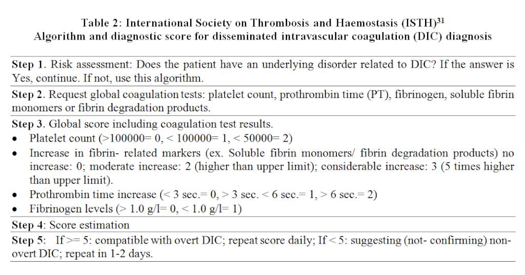
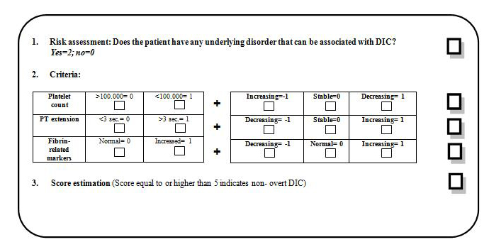
Besides low cardiac output, arterial hypotension and infection there was also a condition compatible with DIC which met Japanese Association for Acute Medicine (JAAM)29 criteria and International Society on Thrombosis and Haemostasis (ISTH)30 “non- overt” DIC criteria. However it did not meet ISTH31 “overt” DIC criteria. High dose vasoactive drugs were also needed for a considerable period. It is surprising that by changing the invasive measurement site for blood pressure it was possible to significantly reduce vasopressor doses.
Our patient was not anticoagulated and did not receive rAPC, both having potentially beneficial effects for DIC and peripheral gangrene.
It can therefore be considered that low cardiac output and sustained hypotension, DIC and vasoactive drugs contributed to SPGS in our patient.
APPENDIX
Alveolar recruitment maneuver used
Having the patient deeply sedated, ventilation shall be applied for 3 minutes under Pressure Control (PC) with an initial 20 cmH2O PEEP and an inspiratory pressure (IP) above 20 cmH2O PEEP. During the first 30 seconds the Vt as well as the hemodynamic tolerance shall be checked. In case de Vt reached is lower than 5 ml/kg (ideal weight) the PC shall be increased by 5 cmH2O. However, if Vt is not reached another 5 cmH2O increase shall be performed to the PC. If hemodynamic tolerance is good then the PEEP shall be increased by 5 cmH2O and then by some 5 cmH2O more until a 30 cmH2O PEEP is reached. By no means should the total pressure exceed 50 cmH2O. Therefore a combination of PEEP (between 20 and 30 cmH2O and PC (between 15 and 25 cmH2O) must be obtained to produce the best oxygenation and the best Vt possible together with the best hemodynamic tolerance. Once the maneuver is finished (2 to 3 minutes later, based on the benefit obtained) the patient shall be ventilated once again in the same manner, however the PEEP should be maintained above the previously used. An assessment shall be conducted comparing SpO2 and PaO2 before and after the maneuver (a patient showing an increase above 10% is considered responder) as well as the improvement in alveolar ventilation (drop in PaCO2 for the same volume per minute).
|
IV. Diagnostic algorithm
SIRS criteria
|
Score
|
|
>3
|
1
|
|
0–2
|
0
|
|
Platelet count (109/L)
|
|
|
<80 or >50% decrease within 24 hrs.
|
3
|
|
>80 and <120 or > 30% decrease within 24 hrs.
|
1
|
|
>120
|
0
|
|
Prothrombin time (patient’s value/ normal value)
|
|
|
>1.2
|
1
|
|
<1.2
|
0
|
|
Fibrin degradation products/fibrinogen (mg/L)
|
|
|
>25
|
3
|
|
>10 and <25
|
1
|
|
<10
|
0
|
|
DIC diagnosis
|
>= 4 points
|
IMAGES
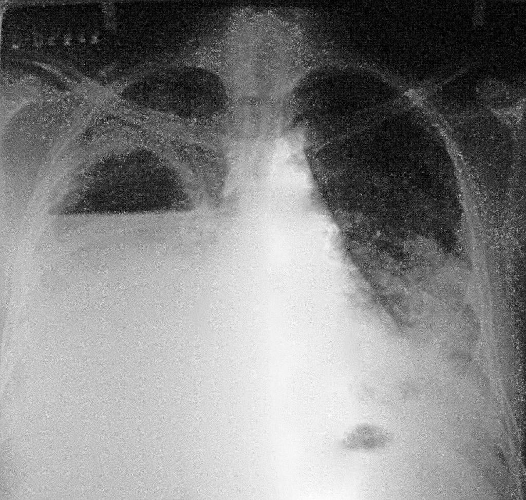
Figure 1: Front chest x- ray: right lung abscess, non- homogeneous left basal pneumonia infiltrate.
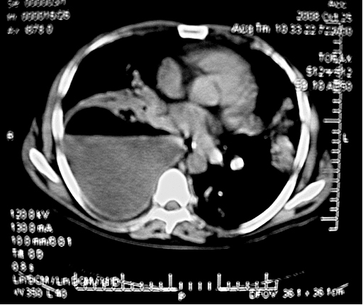
Figure 2: Chest CT scan without IV contrast: right lung abscess, left basal pneumonia infiltrate.
Figure 3: Wet gangrene in right hand. Mid- palm view. Fingers with dry gangrene.
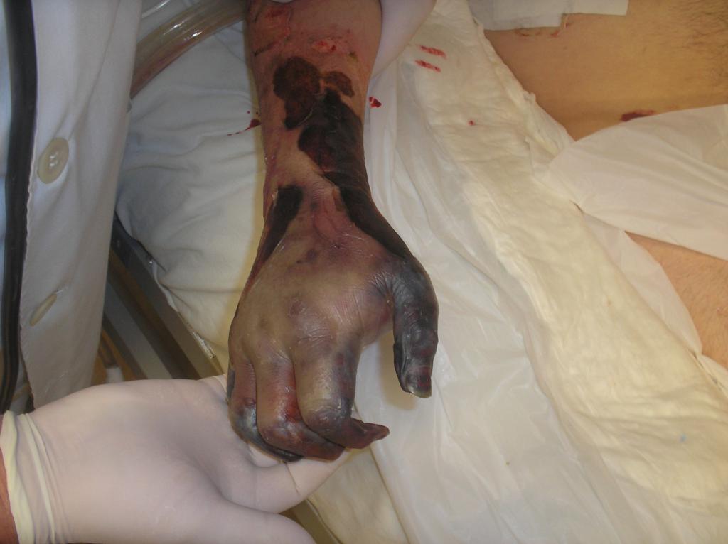
Figure 4: Wet gangrene in right hand. Dorsal view. Dry gangrene in fingers
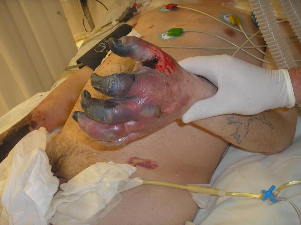
Figure 5: Wet gangrene in left hand. Dorsal view. Dry gangrene in first, second and third fingers
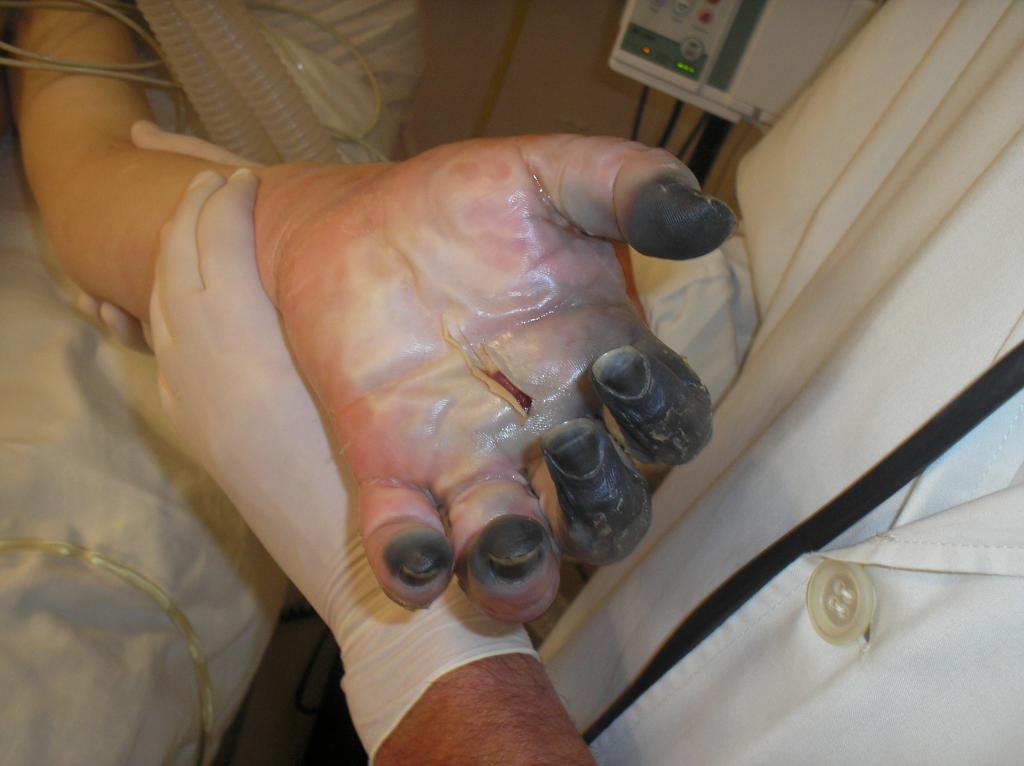
Figure 6: Gangrene in left hand. Palm view. Dry gangrene in fingers.
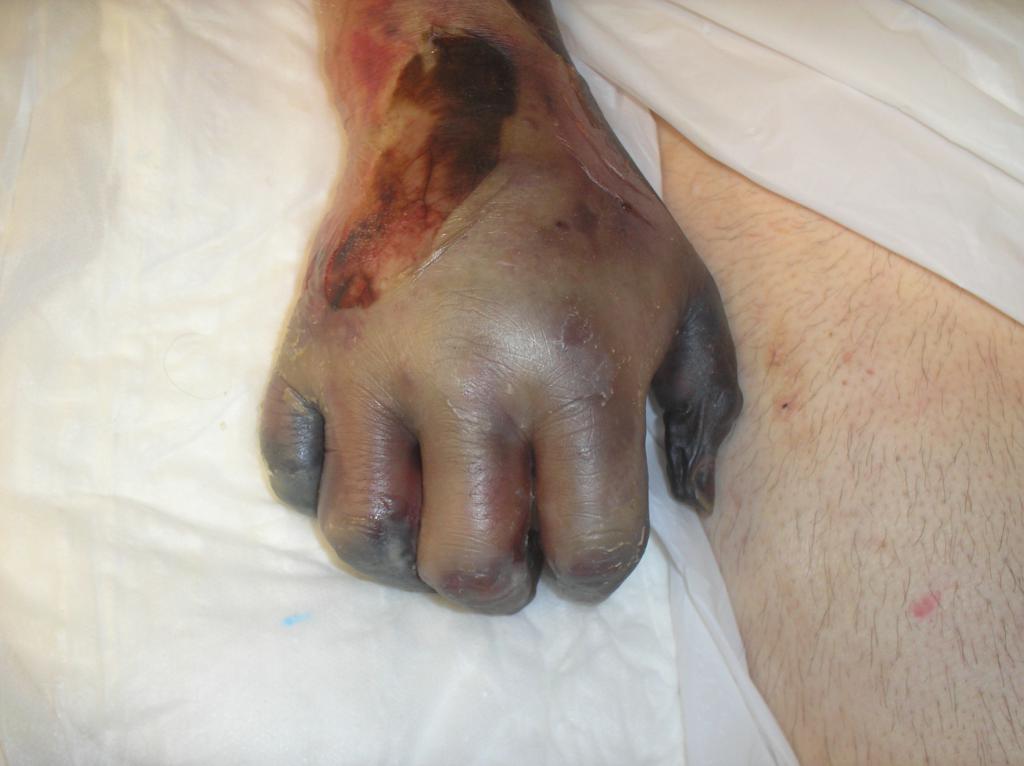
Figure 7: Gangrene in right hand. Dorsal view. Dry gangrene in fingers
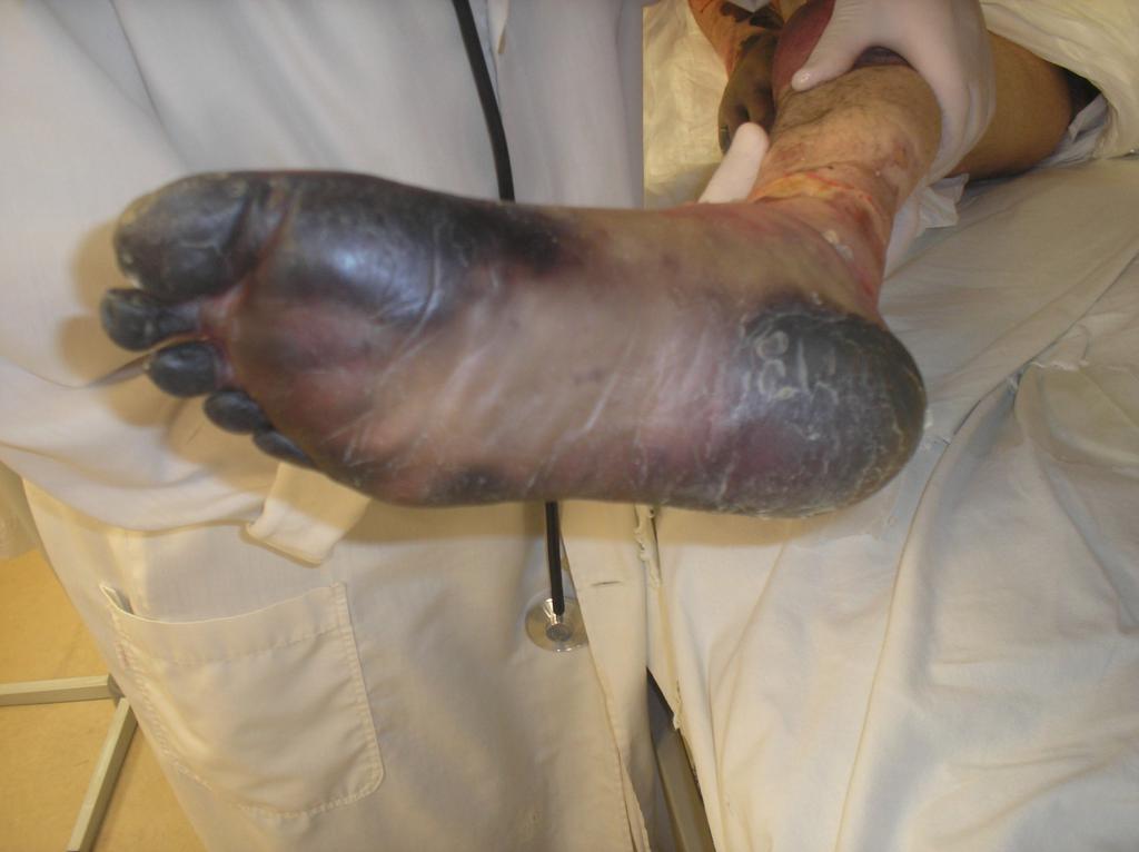
Figure 8: Dry gangrene in right foot. Plantar view. Dry gangrene in plantar area and toes.
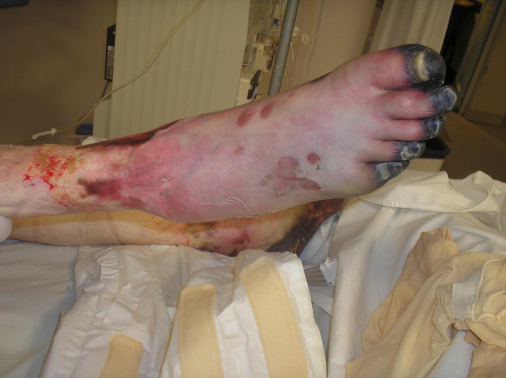
Figure 9: Wet gangrene in right foot. Dorsal view. Dry gangrene in toe
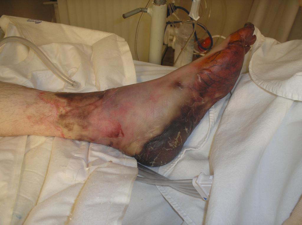
Figure 10: Wet gangrene in left foot. Internal aspect view. Visible thrombosed venous network.
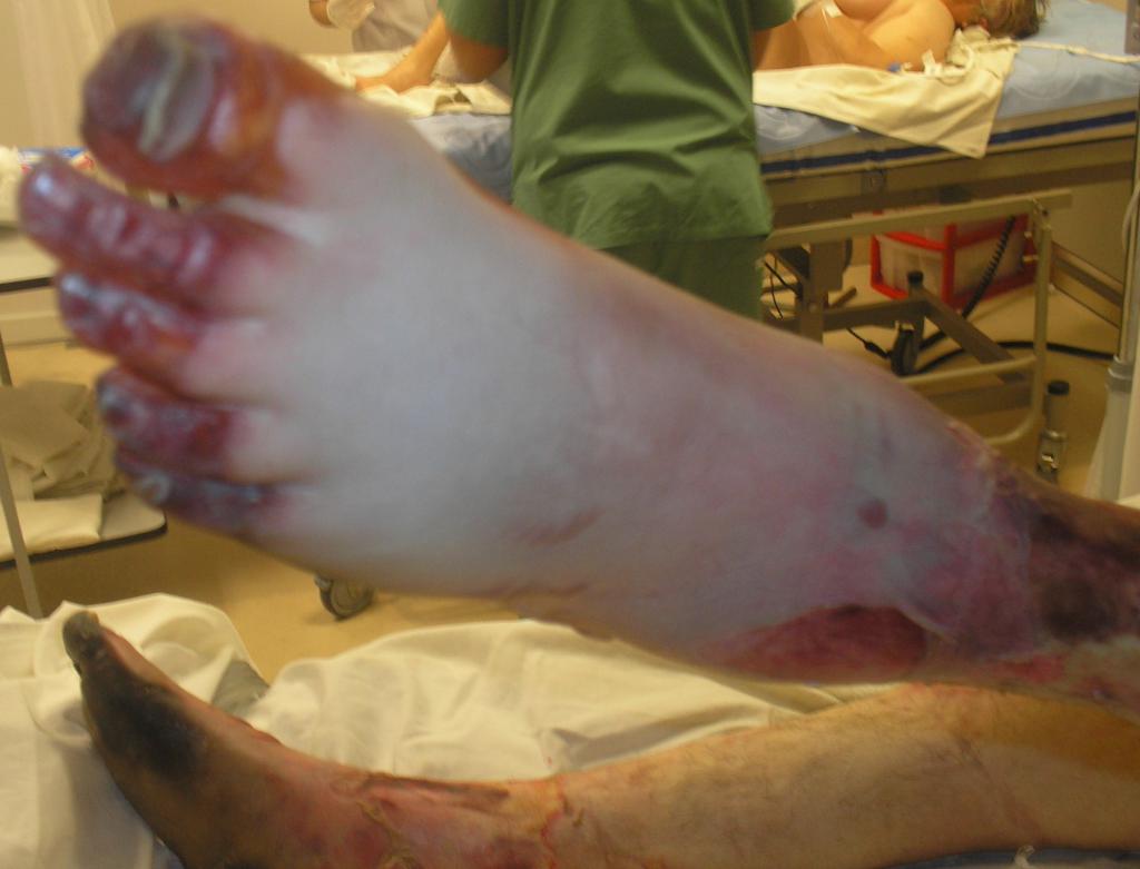
Figure 11: Wet gangrene in left foot. Dorsal view. Dry gangrene in toes
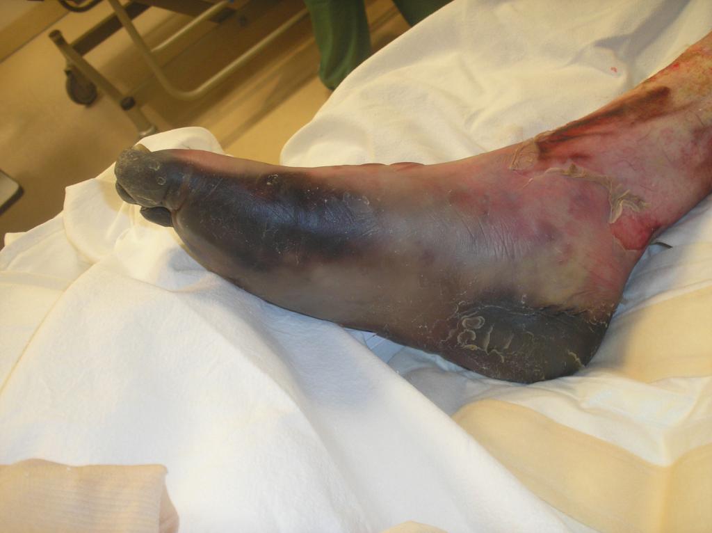
Figure 12: Wet gangrene in right foot. Internal aspect view. Dry gangrene in sole and toes
Competing interests
The authors declare that they have no competing interest.
Consent
Written informed consent was obtained from the patient for publication of this case report and accompanying images. A copy of the written consent is available for review by the Editor-in-Chief of this journal.
Authors contributions
All authors had analyzed and interpreted the patient data, searched and analyzed literature and wrote the manuscript. All authors read and approved the final manuscript.
Acknowledgements
To Donald E Griesdale (MD MPH FRCPC, Assistant Professor, Anesthesiology & Critical Care Medicine, Vancouver General Hospital, University of British Columbia) for his uninterested and kindly academic review and opinion.
REFERENCES
Dellinger RP, Levy MM, Carlet JM, et. al: Surviving Sepsis Campaign: International guidelines for management of severe sepsis and septic shock: 2008 [published correction appears in Crit Care Med 2008; 36:1394-1396]. Crit Care Med 2008; 36:296-327
2 Hernandez G, Peña H, Cornejo G, et al: Impact of emergency intubation on central venous oxygen saturation in critically ill patients: a multicenter observational study. Critical Care 2009, 13:R63.
3 Sud S, Friedrich J, Polli F, et al: Prone ventilation reduces mortalityin patients with acute respiratory failure and severe hypoxemia: systematic review and meta-analysis. Intensive Care Med 2010; 36:585–599.
4 Pelosi P, Gama de Abreu M, Rocco P: New and conventional strategies for lung recruitment in acute respiratory distress syndrome. Critical Care 2010, 14:210.
5 Borges JB, Okamoto VN, Matos GFJ, et al: Reversibility of Lung Collapse and Hypoxemia in Early Acute Respiratory Distress Syndrome. Am J Respir Crit Care Med 2006; 174: 268–278.
6 Kanoore Edul VS, Maskin LP, Dubin A: Effects of recruitment manoeuvres on haemodynamics, oxygen exchange and oxygen delivery in patients with acute lung injury and acute respiratory distress syndrome. Crit Care and Resusc 2010; 12; 143-148.
7 Pestaña D, Royo C, Hernández-Gancedo C, et al: Hemodynamic variations induced by a pressure- volumen curve and an alveolar recruitment maneuver in patients suffering from acute respiratory distress syndrome (Variaciones hemodinamicas inducidas por la realizacion de una curva presion–volumen y una maniobra de reclutamiento alveolar en pacientes con sindrome de distres respiratorio agudo). Rev. Esp. Anestesiol. Reanim. 2008; 55: 348-354.
8 Iannuzzi M, De Sio A, De Robertis E, et al: Different patterns of lung recruitment maneuvers in primary acute respiratory distress syndrome: effects on oxygenation and central hemodynamics. Minerva Anestesiol 2010; 76:692-8.
9 Vincent J.L., Sakker Y, Sprung C, et al.: Sepsis In European intensive care units: Results of the SOAP study. Crit Care Med 2006; 34:344–353.
10 Jansen TC, van Bommel J, Schoonderbeek FJ, et al: Early Lactate-Guided Therapy in Intensive Care Unit Patients. Am J Respir Crit Care Med 2010; 182: 752–761.
1 Bartlett JG: Lung abscess. [http://www.uptodate.com/online/content/topic.do?topicKey=pulm_inf/8395&selectedTitle=1~59&source=search_result].
2 Dorman T, Breslow MJ, Lipsett PA, et al: Radial artery pressure monitoring underestimates central arterial pressure during vasopressor therapy in critically ill surgical patients. Crit Care Med 1998; 26: 1646-1649.
3 Mignini MA, Piacentini E, Dubin A: Peripheral arterial blood pressure monitoring adequately tracks central arterial blood pressure in critically ill patients: an observational study. Critical Care 2006, 10:R43.
4 Galluccio ST, Chapman MJ, Finnis ME: Femoral–radial arterial pressure gradients in critically ill patients. Crit Care Resusc 2009; 11: 34–38.
5 Bernard GR, Vincent JL, Laterre PF, et al: Efficacy and safety of recombinant human activated protein C for severe sepsis. N Engl J Med. 2001; 344(10): 699-709.
6Abraham E, Laterre PF, Garg R et al: Drotrecogin Alfa (Activated) for Adults with Severe Sepsis and a Low Risk of Death. N Engl J Med. 2005; 353:1332-41
7 Vincent JL, Bernard GR, Beale R et al: Drotrecogin alfa (activated) treatment in severe sepsis from the global open-label trial ENHANCE: Further evidence for survival and safety and implications for early treatment. Crit Care Med 2005; 33:2266–2277.
8 Guidelines for the diagnosis and management of disseminated intravascular coagulation. British Journal of Haematology 2009; 145, 24–33.
9 Iba T, Asakura H: Comparison between British and Japanese guidelines for the diagnosis and treatment of disseminated intravascular coagulation. British Journal of Haematology 2010; 149, 451–462.
20 Wada H, Asakura H, Okamoto K, et al: Expert consensus for the treatment of disseminated intravascular coagulation in Japan. Thrombosis Research 2010; 125: 6–11.
2 Marik PE, Pastores SM, Annane D, et al: Recommendations for the diagnosis and management of corticosteroid insufficiency in critically ill adult patients: Consensus statements from an international task force by the American College of Critical Care Medicine. Crit Care Med 2008; 36:1937–1949.
22 Marik PE: Critical Illness-Related Corticosteroid Insufficiency. Chest 2009; 135: 181-193.
23 Hayes M, Yau E, Hinds C, et al.: Symmetrical peripheral gangrene: association with noradrenaline administration. Intensive Care Med 1992; 18:433-436.
24 Davis M, Byrd J, Lior T et al.: Symmetrical Peripheral Gangrene Due to Disseminated Intravascular Coagulation. Arch Dermatol 2001; 137: 139-140.
25 Hutchinson J: Severe symmetrical gangrene of the extremities. BMJ 1891; 2: 8-9.
26 Parmar M.: Symmetrical peripheral gangrene: a rare but dreadful complication of sepsis. CMAJ 2002; 167 (9): 1037-1038.
27 Ghosh SK, Bandyopadhyay D, Ghosh A.: Symmetrical peripheral gangrene: a prospective study of 14 consecutive cases in a tertiary-care hospital in eastern India. J Eur Acad Dermatol Venereol 2009; 23:1–5.
28 Berrey MM, Van Burik JA.: Images in Clinical Medicine: Symmetric Peripheral Gangrene. N Engl J Med. 2001; 344 (21): 1593.
29 Gando S, Saitoh D, Ogura H, et al.: Natural history of disseminated intravascular coagulation diagnosed based on the newly established diagnostic criteria for critically ill patients: Results of a multicenter, prospective survey. Crit Care Med 2008; 36:145–150
30 Toh CH, Downey C.: Performance and prognostic importance of a new clinical and laboratory scoring system for identifying non-overt disseminated intravascular coagulation. Blood Coagulation and Fibrinolysis 2005, 16:69–74.
3 Levi M.: Disseminated Intravascular Coagulation: What’s New?. Crit Care Clin 2005; 21:449–467.