
Date: 24 Feb 2010; Answer available
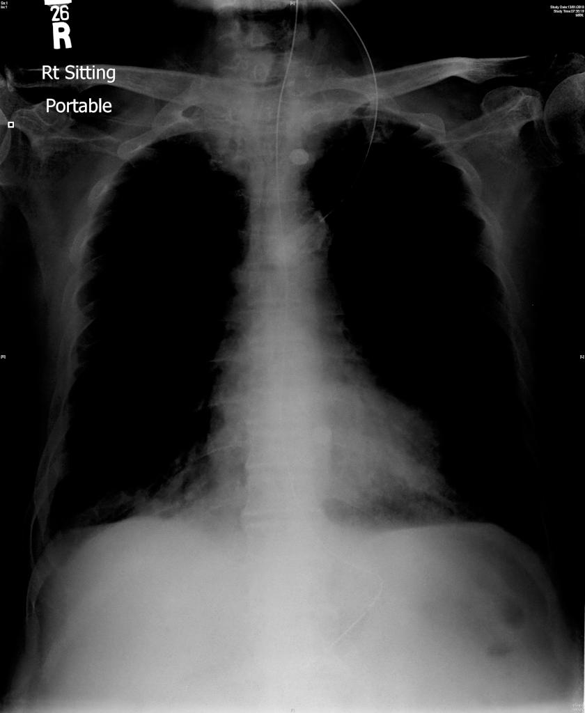
Figure. CXR of the patient (click Read More to see larger image)
The patient was a 59-year-old man with good past health. He was sent to hospital for being found comatose at home by his neighbour. On arrival, SaO2 was barely detectable, and his breathing was noted to be shallow associated with "stidor".
Emergency endotracheal intubation was performed becasuse of impending respiratory arrest. He was then admitted into intensive care unit. First ABG showed pH 7.22; pCO2 72 mmHg; pO2 140 mmHg, HCO3 of 31 and BE at -2. Ventilation with volume cycle at tidal volume 450ml and rate at 16 per minute led to high peak airway pressure of 33, and flow-time curve showed pattern of airflow obstuction.
CXR taken after intubation raised suspicion of tracheal stenosis, as reflected by apparent decrease in diameter of intrathoracic trachea.
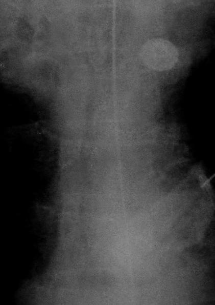
Figure. Magnified view of CXR at upper mediastinum
Plain CT thorax was performed. What sign was shown here?
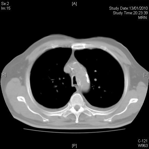
Answer: Saber-sheath trachea deformity
This is pathognomonic for patients with COPD. The sagittal-to-coronal diameter ratio of intrathroacic trachea exceeds 2:1. This conformational change is believed to be related to mechanical force secondary to lung hyperinflation.
Reference
Marom EM, Goodman PC, McAdams HP. Diffuse abnormalities of the Trachea and Main Bronchi. AJR 2001; 176: 713-717.