
Submitted by Dr LEUNG Yuk Wah Natalie, ICU, Pamela Youde Nethersole Eastern Hospital, Hong Kong, on 17 Nov 2009
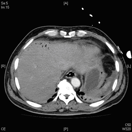
Figure 1. CT abdomen (more images below)
A 54-year old man with good past health was admitted to our hospital due to collapse at home. He developed asystole at ambulance with return of spontaneous circulation after 5 minutes of CPR. The GCS was 3/15 at AED, and both pupils equal and reactive to light. Urgent CT brain did not show any haemorrhage or lesion. He was transferred to the ICU for further care. The best GCS in ICU was E4VTM5, with right side weakness. His condition remained stable until the second day after admission. He developed shock and tachycardia, required fluid and inotropic support. He developed abdominal distension with > 200ml fecal like material aspirated from RT. Blood test showed worsening of metabolic acidosis (pH 7.28, HCO3 21.1 mmol/l, BE -5.6mmol/l). AXR showed dilated small and large bowels, urgent CT abdomen was done (Figures 1 to 4). What are the abnormalities in the CT abdomen? What is your diagnosis? (Click READ MORE for more images)
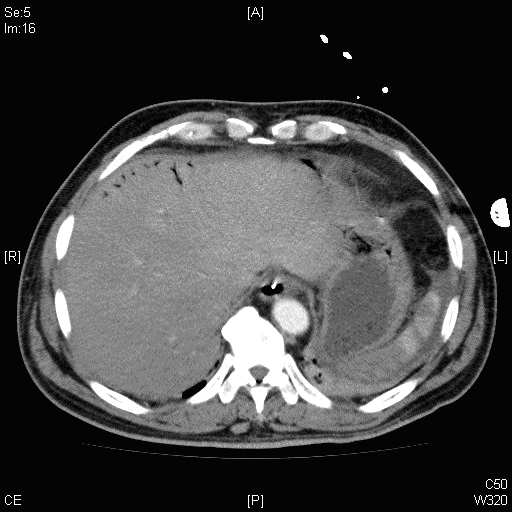
Figure 2
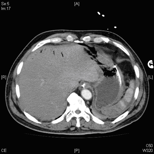
Figure 3
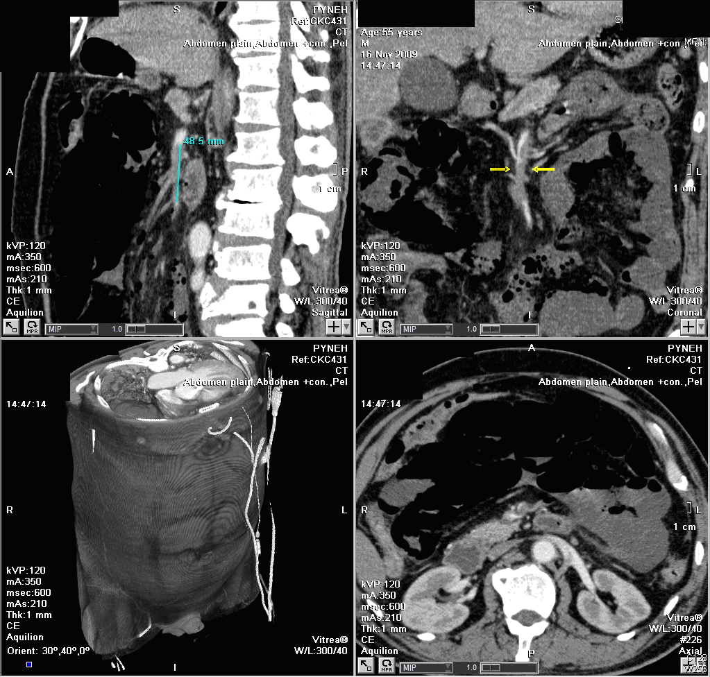
Figure 4
Answers
1. Hepatic portal venous gas
2. Heterogenous enhancement in spleen, probable splenic infarct
3. Filling defect in superior mesenteric artery (SMA)
Acute mesenteric ischaemia due to SMA thrombosis, with extensive hepatic portal venous gas.
Discussion
Hepatic portal venous gas (HPVG) is a rare radiological finding. It is first described in infants in 1955 and then reported in adults in 1960.
Possible causes of HPVG include mesenteric ischaemia, inflammatory bowel diseases, post-procedural complications, colon cancer, gastric ulceration and acute pancreatitis.
The gas in the hepatic portal venous system can be due to either gas under pressure in the bowel lumen or an alteration of the bowel mucosa allowing gas enter the portal system through the mesenteric veins. Increase intraluminal allow air to diffuse out of the bowel lumen, with or without mucosal injury. Mucosal injury result in increased gut permeability to air and allowing intrusion of intraluminal gas into the bowel wall and escape of gas into the portal venous system via the mesenteric veins.
HPVG is not a predictor of mortality. The mortality rate is related to the underlying cause. In a review of 182 cases of HPVG in adults, the overall mortality rate was 39%. The mortality rate was significantly higher at 75% in patients who had bowel ischaemia. On the other hand, the mortality rate was much lower in other conditions like inflammatory bowel disease or intraperitoneal tumor.
In our case, urgent laparotomy was performed which found extensive gangrene of small bowel from 20cm distal to duodeno-jejunal to caecum. He developed rapid deteriorating course and died the next day after operation.
In conclusion, HPVG is a rare radiological finding which warrants immediate attention and requires urgent surgical exploration especially in patients with clinical suspicion of bowel ischaemia.
Reference
1. Chau TN, Loke KL. Leung KS. Hepatic portal venous gas complicating septic thrombophlebitis of the superior mesenteric vein. Hong Kong Medical J 2007; 13: 69-72
2. Gan HN, Tan KY, Chong CK. Finding hepatic portal venous gas in an adult patients: its significance. Singapore Med J 2006; 47(9):814-816
3. RM Calcroft, GM Joynt, J Kew. Ischaemic bowel detected as hepatic portal venous gas on a chest X-ray. Hong Kong Medical J 1999; 5: 303