
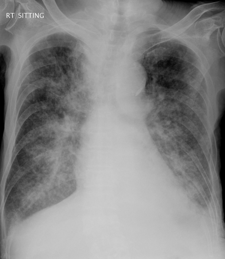
The CXRs showed numerous well-defined non-cavitating non-calcified micronodular shadows in a bilateral diffuse pattern, affecting lower lobes smore than upper. Sizes are variable, and some of them are larger than 5mm, but the largest ones are still < 10mm. Apart from the nodules in lung field, there is reduction of right upper lobe volume as evidenced by rightward deviation of trachea and uplifting of the right horizontal fissure. One should not miss the presence of some right apical homogenous shadow which may represent loculated fluid or mass, and the small left pleural effusion. No mediatinal lymphadenopathy can be appreciated, and bony structures are intact. Anyway, the features are not compatible with typical pneumonia caused by bacteria.
Differential diagnosis for theses microndular shadows are:
(1) Miliary tuberculosis: though the sizes are a bit large
(2) Granulomatous disease: blastomycosis, coccidioidomycosis
(3) Viral pneumonia, in particular VZV
(4) Nocardiosis
(5) Metastasis: broncho-alveolar cell, small cell, thyroid, melanoma, renal cell, lymphoma
If the patient was fit enough, one may subject him to CT thorax and transbronchial lung biopsy. However, he deteriorated rapidly before anything can be delivered. He passed away on day 2 of ICU stay. Paramortem lung biopsy showed carcinomatosis of probable small cell carcinoma from lung primary. Full post-mortem was rejected by family.
This case illustrates the importance of pattern recognition of CXR abnormalities in managing cases with respiratory failure.